- Principles of Fluid Therapy - source
- The principles of therapy are relatively simple; the physical, logistical, and economic constraints can be (and are) overcome by creative, resourceful practitioners. Administration of effective and economical fluid and electrolyte replacement therapy is achievable by every large animal practitioner.
- The most frequent indication for fluid therapy for calves is neonatal calf diarrhea. Regardless of the etiologic organism, the metabolic changes resulting from diarrhea in calves are similar. They include (1) dehydration, (2) acidosis, (3) electrolyte abnormalities, and (4) negative energy balance and/or hypoglycemia. The major cause of dehydration of these calves is fecal fluid loss, which can be as much as 13 per cent of body weight in 24 hours. Compounding this problem is decreased intake from either anorexia or withdrawal of milk by the owner. Acidosis results from bicarbonate and strong cation loss in the stool, lactic acid accumulation in tissues, decreased renal excretion of acid, and increased production of organic acid in the colon in malabsorptive diarrheas. Along with water and bicarbonate, Na, Cl, and K are lost in the feces, which results in a total body deficit of these ions. Negative energy balance can occur in diarrheic calves owing to decreased milk intake, decreased digestion or absorption of nutrients, or replacement of milk with low-energy oral rehydration solutions. Increased energy demand, such as that resulting from cold weather or fever, exacerbates these problems.
- Patient assessment
- The range for PCV in healthy neonatal calves is 22 to 43 per cent, much too variable to provide reliable quantitative information of hydration status, at least with a single sampling. The TPP is even more variable, depending greatly on the degree of colostral immunoglobulin absorption that occurred, as well as hydration. Without a reliable quantitative measure for hydration status, we must rely on estimates based on clinical signs. Table -1 provides a guideline for estimating the degree of dehydration in cattle. This table is based on research conducted by Constable et al,1 and is the most critically validated estimate of dehydration in calves. However, even in the absence of a validated system of estimating degree of dehydration, rehydration has been clinically successful, suggesting that precise estimates are not necessary. Rather than becoming overly concerned with pinpointing the exact degree of dehydration, veterinarians should be concerned whether intravenous therapy is needed, or whether voluntary or forced oral supplementation will suffice. Rather than defining an exact long-term fixed plan for rehydration, we should begin with a reasonable plan and adjust it as needed. In other words, guess and reassess.
- Table 1: Guide to estimation of fluid replacement requirement
- Data from Constable PD et al: Use of hypertonic saline-dextran solution to resuscitate hypovolemic calves with diarrhea. Am J Vet Res 57: 97-104, 1996
- Empirically, 8 per cent dehydration is the severity beyond which it is considered that oral fluid therapy will not suffice. According to the table above, 8% dehydration is characterized by eyeball recession of 4 mm in the skin tent duration of 6 seconds. Other clinical signs associated with severe dehydration include dry mucous membranes, and moderate to severe depression. Calves displaying these signs will benefit the most from intravenous therapy. In general, calves that readily suckle quantities of rehydration solution sufficient to meet their replacement, maintenance, and ongoing loss needs will respond to oral solutions. Many of the more severely dehydrated calves will respond to forced oral solutions as well, but intravenous replacement is preferred.
- Acidemia can quickly and accurately be assessed when a blood gas analyzer is available. Measurement and assessment of total carbon dioxide (TCO2) will provide essentially equivalent clinical data in assessment of non-respiratory acidosis or alkalosis, which is the type of acid-base disturbance most frequently encountered in conscious animals. In most cases in practice, the degree of acidosis will be estimated. Naylor determined that dehydrated calves greater than 1 week of age had more severe acidosis (mean base deficit of 19.5 mEq/L) than did those less than 1 week of age (mean deficit of 14.4 mEq/L). As a rule of thumb, severely diarrheic calves less than 1 week of age can be assumed to have a base deficit of 10 to 15 mEq/L whereas those greater than 1 week of age can be assumed to have a base deficit of 15 to 20 mEq/L.
- Laboratory analysis of serum or plasma electrolytes can be of benefit in evaluating the replacement needs of diarrheic calves but, if misinterpreted, could lead to inappropriate therapy. Plasma represents a very small portion of total body water, and the concentration of electrolytes in a blood sample must be interpreted in light of that fact. If Na and Cl are within normal limits and a calf has lost 20 per cent of ECF volume, then the calf has a total body Na and Cl deficit of nearly 20%. Because Na and Cl concentrations are often within or below the reference range in diarrheic calves, it is extremely important to provide these electrolytes in replacement solutions. Failure to do so will result in dilution of the already deficient ions.
- A potentially more misleading laboratory value than plasma Na and Cl is plasma K. Many dehydrated, acidemic calves are hyperkalemic, yet they have a total body K deficit. This paradox is the result of a shift of K out of the ICF compartment into the ECF compartment during acidemia. The ECF, which normally contains only about 5 per cent of the body’s total exchangeable K, has a greater than normal concentration of K, however, ICF K concentration and total body K content are decreased.
- Blood glucose determination can be made by a serum analyzer or by a rapid method using a hand-held meter. Hypoglycemia in calves results in weakness, lethargy, coma, convulsions, and opisthotonos. Negative energy balance is not easily quantified because it can result from inadequate intake, malabsorption-maldigestion, or increased metabolic demand due to fever or low ambient temperature.
- Estimating fluid and electrolyte replacement requirements
- The first priority in treatment of a dehydrated calf should be restoration of ECF volume. When estimating the volume of fluid needed by a patient, the veterinarian should consider not only the deficit, but also maintenance requirements (50 to 100 ml/kg) and compensation for continuing loss (as much as 4 L in 24 hours). In many cases, the actual deficit is less than half of the total 24-hour volume requirement.
- Second in priority to correcting ECF volume depletion is correcting acidosis. It has been
- suggested that the restoration of ECF volume alone would allow the kidneys to eliminate acid in sufficient quantity to restore normal acid-base balance. In most cases with baby calves it is necessary to treat the calf rapidly and, in beef calves, return them to the care of its mother as soon as possible. The ability of non-alkalizing fluids to rapidly correct moderate to severe academia in calves has been disproved. Neither intravenously or orally administered solutions without alkalinizing agents resolved acidosis expeditiously even through ECF volume was restored.
- Acidosis can be corrected by the administration of bicarbonate ions or so-called bicarbonate precursors, salts of weak organic acids (lactate, acetate, gluconate). Studies in calves have demonstrated the superior rapid alkalinizing efficiency of bicarbonate, compared with L-lactate and acetate. Sodium bicarbonate is the most economical and readily available alkalinizing agent; however, it cannot be heat sterilized. It also should not be used in solutions containing calcium because an insoluble compound will form.
- Alternative alkalinizing agents offer both advantages and disadvantages. Hepatic perfusion and function are necessary for lactate metabolism, and endogenous lactate (lactic acid) that accumulates during hypovolemia and shock can reduce its metabolism. Acetate has the advantage of being metabolized by peripheral tissues and of having no significant endogenous source and no unmetabolized isomer. Gluconate, an alkalinizing agent used in combination with acetate in some commercially-prepared solutions for intravenous administration to people, dogs, and horses, has been shown to be ineffective as an alkalinizing agent in calves when administered intravenously.
- Rate of administration of alkalinizing agents, especially sodium bicarbonate, is a controversial subject. Whereas numerous warnings about CSF acidosis can be found in veterinary literature, the author is not aware of a documented clinical case of CSF acidosis in domestic animals and therefore does not hesitate to replace the total calculated deficit of bicarbonate in the initial deficit replacement solution.
- When blood gas analysis is available, the value for the base deficit (BD) can be used to calculate total base requirement by use of the formula.
- •0.6 X BD X body weight = base requirement in mEq
- When the value for TCO2 or bicarbonate is known, it can be subtracted from 25 (the approximate normal value for plasma bicarbonate), and the difference can be used in place of BD in the formula. When it is not possible to quantify acid-base status, an estimate of 10 to 20 mEq/L may be used to formulate fluids for intravenous use for diarrheic calves. Remember that calves greater than 1 week of age tend to become more severely acidotic.
- Glucose stimulates the release of insulin, which in turn enhances the movement of K from the ECF to the ICF. It also provides readily available energy. Glucose concentrations of 1 to 2 per cent in intravenously administered solutions have produced favorable clinical results and usually do not result in significant glucosuria or osmotic diuresis. In selected cases, total or partial parenteral nutrition may be beneficial to calves with severe prolonged malabsorptive diarrhea.
- The importance of replacing Na and Cl should not be overlooked. Remember that total body Na and Cl are deficient in dehydrated calves, even when plasma concentrations are normal.
- The administration of K to hyperkalemic acidemic calves can be accomplished safely if HCO3 and dextrose are administered concurrently. As previously mentioned, dextrose and HCO3 enhance the movement of K from the ECF to the ICF. Ideally, the initial liter or so of intravenously administered rehydration solution should contain less K than subsequent volumes. However, practicality often dictates the use of a single solution for rehydration. There seems to be little danger in including up to 20 mEq of K per liter if bicarbonate and dextrose are included in the solution.
- Formulating a solution for intravenous administration
- There are as many correct ways to formulate solutions for intravenous administration in calves as one can imagine. The following is a list of suggested criteria for intravenously administered solutions.
- 1. Osmolality between 300 and 450 mOsm/L.
- 2. Na and Cl concentrations near 140 and 100 mEq/L, respectively.
- 3. K concentration 10 to 20 mEq/L. (Because 1 g of KCl contains 14 mEq K, inclusion of 1 g of KCl/L fulfills this criterion.)
- 4. Dextrose at 10 to 20 g/L of solution (1 to 2 %).
- 5. NaHCO3 or a suitable metabolizable base calculated to meet the measured deficit (or an estimated BD of 10 to 20 mEq/L if laboratory values are not available).
- Of course, commercial solutions like lactated Ringer’s can be used. In most cases, NaHCO3 will be required in addition to correct acidosis. Dextrose and additional K should also be added. Remember that HCO3 should not be mixed in the same container with calcium-containing solutions, such as lactated Ringer’s. Therefore, one strategy utilizing commercial solutions is to administer NaHCO3 solution initially (2 L of 1.3% NaHCO3 or 0.5 L of 5% NaHCO3 solution) followed by Lactate Ringer’s solution with added K and dextrose.
- Whereas it may be ideal to rehydrate a patient over 24 to 48 hours, bovine practitioners must often use the maximal safe infusion rate rather than the ideal. A maximum of 80 ml/kg/hr has been suggested as a safe flow rate for calves. A more conservative rate of 50 ml/kg/hr is probably a reasonable, relatively safe maximal infusion rate. With use of this infusion rate, most calves can be rehydrated in 2 to 3 hours. During rapid intravenous administration of fluids, the veterinarian or attendant should periodically monitor heart rate, respiratory rate and character, and attitude, adjusting the flow rate if necessary. Extra caution should be exercised when administering intravenous fluids to hypothermic calves.
- When possible, it is desirable to administer approximately 1 L of the solution rapidly to reverse hypovolemic shock, and then administer the balance over a period of hours. This will maximize the benefit of the therapy by minimizing the diuresis that is sometimes induced by rapid fluid administration. If it is impractical or impossible to administer the total 24-hour requirement, or even the total deficit by intravenous infusion, 1 or 2 L of fluid administered intravenously may be enough to improve the circulatory status of a calf so that the balance of the calf’s requirement may be provided by the oral route. In other words, a relatively small volume of fluids administered intravenously may convert a calf from the intravenous fluid required to the oral fluid satisfactory category. Fluids for maintenance and continued loss may be administered orally or by slow intravenous infusion. Alternatively, intravenously administered hyperosmolar saline solution combined with intraruminally administered electrolyte solution may be administered.
- Hypertonic saline solution (HSS)
- Studies reveal that intravenous and infusion of HSS plus ORS is essentially equivalent to large volume isotonic fluids for treatment of dehydration and shock in calves. HSS restores cardiovascular function and perfusion rapidly, while ORS addresses the acidosis, energy deficits and K deficits. HSS 7.2% should be administered at a dose of 4 ml/kg over 3-10 minutes. Rapid administration is essential because its effect is caused by transiently creating intravascular hyperosmolality. The effect of HSS is short-lived. Colloids such as dextran and hetastarch, enhance the efficacy of HSS by prolonging its effect, but they add substantially to its cost.
- Placement of an auricular vein catheter is simpler but requires a smaller catheter, therefore limiting the flow rate of the fluids. In most cases, however, the flow rate is sufficient for rehydration. The dorsal ear is clipped if necessary and aseptically prepared. A tourniquet (rubber band) is stretched around the base of the ear around the ear dorsally to ventrally and using a hemostat clamped on the ventral aspect of the year. Any visible vein that is sufficiently long and straight is acceptable. It is very important however to introduce a catheter as far from the base of the ear as possible so that the catheter tip does not extend to the base of the ear causing it to bend and occlude when the ears move. After introduction and placement, the catheter is capped and flushed with flushed with heparinized saline and superglue is applied. A light tape bandage is applied to secure the catheter to the ear.
- Fluid therapy for mature ruminants
- Much is known and much has been written about fluid therapy for calves. Most dehydrated calves are become dehydrated because of diarrhea. A relatively consistent blood gas and electrolyte profile is produced. One of the major metabolic derangements is acidosis. Many commercial oral preparations are available. It is necessary to discuss fluid therapy of mature cattle separately from fluid therapy of calves because the metabolic abnormalities commonly seen in mature cattle are quite different than those of calves and other species. Few commercially prepared solutions are appropriate for mature cattle with alkalosis. While neonatal calves tend to become acidotic mature cattle tend to become alkalotic. The exceptions are discussed below. The purpose of the paper is to provide guidelines for fluid therapy strategies for mature cattle without the use of extensive laboratory data.
- Diseases that consistently result in metabolic acidosis in mature cattle include carbohydrate engorgement, and esophageal obstruction or dysphagia. This is due to excessive fermentation and acid build-up in carbohydrate engorgement and salivary bicarbonate loss in esophageal obstruction and dysphagia. Other less common and less consistent causes include diarrhea, pneumonia, fatty liver disease, urinary tract disease and late-stage abomasal volvulus. In a study of over 600 cattle, when those with pneumonia, urinary tract disease and diarrhea were excluded, alkalinizing fluids were indicated in only 16% of dehydrated mature cattle. Aggressive, alkalinizing therapy should be administered without laboratory assessment of acid-base status only in carbohydrate engorgement and esophageal obstruction.
- Hypochloridemia and hypokalemia were relatively common electrolyte abnormalities, occurring in nearly 50% and 25% of mature cattle, respectively. These trends were stronger in dairy cattle. These abnormalities were most frequently seen accompanying alkalosis. Elevated electrolyte values were uncommon in this study.
- Fluid therapy for mature cattle with acidosis
- In mild cases of carbohydrate engorgement, oral therapy may suffice. Because ruminal and systemic acidosis are present, alkalinizing agents like magnesium hydroxide are indicated. Additional volume can be supplied by pumping ruminant electrolyte solution, saline solution or tap water intraruminally. Remember, if the rumen is distended with fluid, do not administer oral fluids. There is already an excess of fluid in the rumen in the face of dehydration. Adding more will only increase abdominal distention. The excess fluid should be removed if possible, the rumen alkalinized and intravenous fluid therapy initiated. Glucose-containing solutions (like commercial calf electrolytes) are probably not indicated because they may enhance fermentation and acid production. In order to determine the amount of base to administer, the same formula as for the calf is used except 0.3 is used as the factor for ECF rather than 0.6 :
- • Bicarbonate deficit (mEq) = Base deficit (mEq/L) x 0.3 x Body weight (kg).
- For a 600 kg cow with an estimated base deficit of 15 mEq/L (moderate acidosis), we obtain a deficit of 2700 mEq. If we use 8.3% NaHCO3 solution, 3 L is required. If we use 5% NaHCO3 solution 4.5 L is required. If we use isotonic NaHCO3 solution, 18 liters is required to correct the acidosis.
- Fluid therapy for cattle with alkalosis
- Most gastrointestinal diseases of cattle and many other causes of dehydration (with the exception those mentioned above) are associated with metabolic alkalosis, hypokalemia and hypochloridemia. The degree of these changes may be mild (as in early left displaced abomasum) or severe (as in abomasus volvulus). If the gastrointestinal tract is patent and functional, oral rehydration is the method of choice. If obstruction, ileus or circulatory shock is present, intravenous fluids should be administered. When isotonic solutions are used, they should contain sodium in approximately physiologic concentrations, while potassium and chloride should be provided in concentrations greater than that of plasma. For dairy cattle, I like to add calcium as well. We use our own ruminant electrolyte solution as a routine fluid for oral or IV administration. (Table 1) Usually if we place a catheter intravenously, we administer at least 20 liters IV. At least half of the total deficit should be given IV. The balance of the fluid deficit can be corrected by oral administration if the G.I. tract is functional.
- Dextrose may be added to intravenous solutions for anorectic cows or those with ketosis. Each 500 ml of 50 % dextrose will add 1.5 % glucose to 20 liters of solution. Up to four 500 ml bottles (yielding a 5% solution) can be added, although I prefer 2.5 %. Alternatively, hypertonic saline (7.2% NaCl) solution may be used to correct dehydration and electrolyte abnormalities with smaller volumes of IV fluid and shorter infusion times as long as water is provided either by free choice or forced administration. For a 500 kg animal, the dose is 2 liters. The end result is very similar compared to IV administration of large volumes of isotonic fluid
- Remember, alkalinizing solutions (those containing lactate, acetate, or bicarbonate in greater than physiologic concentrations) are seldom needed in adult cattle except in those cases mentioned on the previous section. Oral solutions formulated for calves often contain: 1) NaHCO3, which is contraindicated in ruminal alkalosis (frequently seen in anorectic cattle) 2) glucose, which will be rapidly consumed by ruminal microbes. Therefore, they are expensive and less than ideal for mature cattle.
- References
- Roussel AJ, Cohen ND, Holland PS, Taliaoferro L, Green RA, enson PA, Navarre CB (1998), Alterations in acid-base balance and serum electrolyte concentrations in cattle: 632 cases (1984-1994)., Journal of the American Veterinary Medical Association 212: 1769-1775
- Roussel AJ, Taliaoferro L, Navarre CB, Hooper RN (1996), Catheterization of the auricular vein in cattle in cattle:68 cases, Journal of the American Veterinary Medical Association 208: 905-907
- Roussel AJ (1990), Fluid therapy in mature cattle, Vet Clin North Am Food Anim Pract 6(1): 111-123
- Maintenance Fluid Plan in Animals - source
-
Maintaining intravascular fluids after resuscitation from hypovolemic shock and during systemic inflammatory response syndrome disease conditions causing increased capillary permeability can be a challenge. Hydroxyethyl starch solutions can be administered as a constant-rate infusion at 0.5–1 mL/kg/hour in dogs, or 0.25–1 mL/kg/hour in cats. Newer HES solutions may be administered at higher rates (2 mL/kg/hour) without impacting coagulation. The dosage is adjusted to maintain an adequate mean arterial pressure and CVP. The amount of crystalloids administered with colloids must be reduced by 40%–60% of what would be administered if crystalloids were used alone.
-
The maintenance fluid plan should address three ongoing requirements: replacement of lost interstitial volume (rehydration), maintenance fluids (for normal homeostasis), and replacement of ongoing losses. The volume of rehydration fluids required is determined by reassessing hydration parameters after resuscitation, using the following formula: % dehydration × body wt (kg) × total body water (0.6). This volume is commonly administered throughout 4–12 hours with standard isotonic, balanced electrolyte replacement fluids.
-
Maintenance fluid requirements are added to the rehydration rate. Maintenance fluids are calculated with one of the following formulas:
30 x body weight (kg) + 70 = mL of maintenance fluids per 24 hours
(70 x body weight (kg))0.75 = maintenance fluids per 24 hours
-
The second formula is necessary for patients <2 kg or >70 kg. With prolonged parenteral fluid administration, usually throughout a course of days, serum sodium may increase, and maintenance fluids (eg, half-strength saline or 5% dextrose in water) may be needed to replace free water deficits.
-
Ongoing or increased fluid losses vary substantially and must be estimated and replaced. Ongoing losses can be estimated by measuring urine and fecal output, nasogastric tube suction, bandage weight, or vomitus volume. Patients should be weighed regularly to determine changes in body weight. Insensible losses, which can be increased with fever, wounds, higher metabolic demands, and other factors can increase the maintenance rate by 15–20 mL/kg/day. Increased fluid requirements in patients with fever have not been well established in veterinary medicine; in human medicine, an increase 1°C (1.8°F) above normal body temperature may increase fluid requirements by 10%–12%.
-
Monitoring Fluid Therapy of Maintenance Fluid Plan in Animals
- All animals receiving fluids should have a physical examination, including assessment of hydration and body weight, with urine production checked at least twice per day, more frequently in the critically ill. Overzealous administration of crystalloids can manifest as increased respiratory rate and effort, crackles or wheezes on auscultation, serous discharge from the nares, chemosis, jugular vein distention or pulsations, shivering, edema, hypertension (>140–150 mmHg systolic), increased CVP (>8–10 cm H2O), significant increase in body weight (>12%–15%), and rapid and/or dramatic decrease in PCV and total solids. In animals with urinary catheters, urine output can be monitored and compared with fluid administration volumes. Monitoring CVP, pulmonary capillary wedge pressures, and cardiac output variables may be helpful in selected animals, although pulmonary artery catheters are rarely placed. Monitoring electrolytes and PCV/total solids may provide an objective measurement of fluid balance.
- Patients should be regularly monitored for complications associated with fluid administration, which may be grouped by category:
- Delivery method:
- catheter-associated phlebitis
- catheter-related thrombosis
- catheter-related sepsis and fever
- extravasation of fluids
- catheter-related foreign bodies (rare)
- Incorrect fluid prescription:
- fluid intolerance (overload), which may result in ascites, edema, etc
- insufficient fluid administration, which may result in dehydration or shock
- exacerbation of noncompressible hemorrhage
- exacerbation of acid-base abnormalities
- exacerbation of electrolyte abnormalities
- exacerbation of tonicity abnormalities
- development or exacerbation of renal injury (colloids)
- exacerbation of coagulopathies
- exacerbation of anemia
- hyperglycemia (with dextrose-containing fluids)
- When parenteral fluid administration is to be discontinued, the animal should be able to maintain hydration by voluntary drinking and eating or tolerate enteral supplementation (through a feeding tube) or subcutaneous fluid administration. Tapering the volume infused IV throughout 24–48 hours allows the renal medulla to reestablish the osmotic gradient and helps prevent excessive fluid loss through diuresis.
-
Key Points
- There are three categories of shock: hypovolemic, cardiogenic, and distributive.
- The ability to create an effective fluid plan depends on understanding the different body fluid compartments and dynamics.
- There are three major fluid compartments, the intravascular, extravascular, and interstitial spaces, separated by a capillary membrane and an endothelial glycocalyx layer.
- Crystalloids are water-based solutions with small-molecular-weight particles, freely permeable to the capillary membrane, and are interstitial volume replacement solutions.
- Colloids are water-based solutions with a molecular weight too large to cross the capillary membrane and are intravascular volume replacement solutions.
- Determination of appropriate resuscitation endpoints and resuscitation technique is vital to creating a fluid resuscitation plan.
- Patients should be regularly assessed for fluid requirements and complications.
- Patients not responding to IV fluid resuscitation should be assessed for reasons for poor response.
-
- Deterimination of the Fluid Deficit - source
- Loss of fluid volume from the intravascular fluid compartment is manifested by poor perfusion (shock) and inadequate tissue oxygenation. This volume deficit results in a lower vessel wall tension. Decreased wall tension in the aortic arch and carotid arteries results in decreased stimulation of the baroreceptors. This decreased rate of firing, sent via the glossopharyngeal and vagus nerves to the medulla oblongata, results in decreased inhibition (stimulation) of the sympathetic system. Stimulation of the sympathetic nervous system is manifested by clinical changes in heart rate, pulse intensity, blood pressure, capillary refill time, mucous membrane color, level of consciousness, and rectal temperature. These physical perfusion parameters, combined with blood pressure, are used clinically to detect intravascular volume deficits. Most animals with an intravascular deficit (poor perfusion) also have concurrent extravascular (interstitial and intracellular) deficits.
- Fluid deficit in the interstitial and intracellular spaces causes clinical signs of dehydration. Physical findings are used to estimate the percentage of dehydration. Semidry oral mucous membranes, normal skin turgor, and eyes maintaining normal moisture indicate 4%–5% dehydration. Dry oral mucous membranes, mild loss of skin turgor, and eyes still moist indicate 6%–7% dehydration. As dehydration becomes more severe, significant quantities of fluid shift from the intravascular space into the interstitium, causing concurrent perfusion deficits with dehydration. Dry mucous membranes, considerable loss of skin turgor, retracted eyes, acute weight loss, and weak rapid pulses (concurrent intravascular deficit) indicate 8%–10% dehydration. Very dry oral mucous membranes, complete loss of skin turgor, severe retraction of the eyes, dull eyes, possible alteration of consciousness, acute weight loss, and thready, weak pulses indicate ≥12% dehydration.
- The physical guidelines to estimate dehydration are misleading in two common clinical situations. Chronically emaciated and geriatric animals may have metabolized the fat from around the eyes and the collagen in the skin, resulting in poor skin turgor and sunken eyes despite normal hydration. Animals with rapid fluid loss into a third body fluid space (a space within the body cavity where fluid from the local interstitial and intravascular spaces leak) have rapid fluid shifts from the intravascular compartments into these spaces before clinical evidence of interstitial fluid loss is seen. Both situations require evaluation of mucous membrane and eye moisture, PCV, and total solids before dehydration can be estimated.
- Selection of Fluids
- Fluids must be administered that will concentrate within the body fluid compartment where the volume deficit lies. Crystalloids are water-based solutions with small-molecular-weight particles, freely permeable to the capillary membrane. Colloids are water-based solutions with a molecular weight too large to freely pass across the capillary membrane. Colloids are thought of as intravascular volume replacement solutions, and crystalloids as interstitial volume replacement solutions.
- Dehydration in Calves - Assessing Hydration Status:
- It is essential that any animal, particularly calves with diarrhea, have oral, subcutaneous (SQ), or intravenous (IV) fluid supplementation.
- Use the following guidelines to determine the hydration status of an animal:
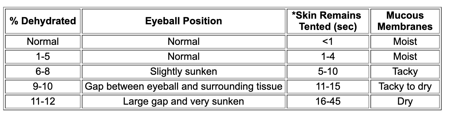
- *See page B115 for additional information on skin tent.
- Fluid Replacement:
- Once the level of dehydration in the calf has been determined, a simple calculation can be done to identify how much fluid is required.
- Take the % dehydrated and multiply by the calf’s body weight in kg.
- For example, an 80 lb. (36 kg) calf is determined to be 9% dehydrated.
- .09 X 36 kg = 3.24 – This means that 3.24 liters of fluid (oral electrolytes, SQ, or IV fluid) are required to replace what has been lost.
- After the initial calculated fluid dose is administered, additional fluids can be given at a rate of 5 mL for every 2.2 lbs., every hour. In addition to the 5 mL/2.2 lbs/hour, an additional 1-4 liters each day may be required to keep up with continued fluid losses because of diarrhea, etc.
- Hints:
- 1 gallon = 3.7 liters and 1 kg = 2.2 lbs.
- In this example the calf weighs 36 kg (80 lbs. divided by 2.2 = 36 kg)
- This fluid can be given orally (PO), SQ, or IV (V is much better in an animal that is down and lethargic). Only sterile fluid (0.9% saline solution or sodium chloride) should be administered SQ or IV. See page F755 for additional suggestions.
- **Oral Fluid Administration: **Oral fluids should only be used in calves that are 1-5% dehydrated. If the calf is more dehydrated than this, oral fluids alone do not meet the calf’s needs. The most common problems with oral fluid administration is that the method and frequency of administration is not ideal and the quantity administered is often insufficient. To overcome these problems, the following suggestions are a must.
- Reconstitute the oral electrolyte exactly according to the manufacturer’s
- instructions.
- Calculate the calf’s fluid requirements using the previous example.
- Administer no more than 2 liters at one time.
- The best way for a calf to obtain oral fluids is by suckling a nipple bottle. This allows the fluid to enter the abomasum through the esophageal groove. If the calf is too weak to suckle, fluids can be administered with an esophageal feeder. See page B850 for instructions on using an esophageal feeder. This method, however, causes the fluid to enter the rumen directly and not the abomasum. Frequent feedings with an esophageal feeder can cause decreased gastrointestinal (rumen/intestines) function and other complications.
- Do not be reluctant to administer milk replacers in addition to electrolytes. Electrolytes do not contain adequate nutrition for the calf and therefore milk replacers are necessary. It is true that some milk replacers can increase the amount of diarrhea that is seen. This is a necessary trade off for the extra nutrition the calf requires.
- All electrolytes and milk replacers should be fed at least 30 minutes apart and never mix the two fluids. Milk digestion is slowed when combined with oral electrolytes.
- The frequency of feedings can be variable and depend on the severity of the fluid loss and management issues. In general, calves that are only slightly dehydrated may require only two oral feedings, while calves that are around 5% dehydrated may require feeding every 2 hours.
- Subcutaneous (SQ) Fluid Administration:This method of fluid administration should be used in those calves that are 6-8% dehydrated. If SQ fluids are given, remember the following:
- If the calf is greater than 8% dehydrated, IV fluids should be given.
- Warm the fluids to body temperature before administration.
- Use only sterile isotonic fluids (0.9% saline solution).
- Because of potential infections, do not add glucose to any fluid given SQ.
- The loose skin areas of the neck, shoulder, and behind the elbow are great areas to administer SQ fluid. The injection area should be cleaned and sterilized before inserting the needle.
- The fluid can be given as one large bolus or can be given over a period of time. Do not give over 500 mL in one location and over 2 liters total to one calf at one time. It usually takes about 4-6 hours for the fluid to completely absorb.
- ***** Oral and SQ fluids can be given at the same time and are often a great method of restoring proper hydration.
- **Intravenous (IV) Fluid Administration: **IV fluid administration requires that a sterile catheter be placed in the animal. This requires professional help and additional training. Once the catheter is in place, the following should be considered:
- Only administer sterile fluids (0.9% saline solution).
- Calculate the animal’s fluid needs by utilizing the table and example at the front of this discussion.
- Most calves are acidotic (meaning that they have a low blood pH). Because of this, bicarbonate should be added to each liter of fluid. If 0.9% sterile saline solution or sodium chloride is used, approximately 150 mL of 8.4% sodium bicarbonate should be added to the first 2-3 liters of IV fluids.
- Supplemental glucose may also be necessary, particularly if the animal is not eating. Placing about 60 mL of a 50% dextrose solution in each
- 1 liter of IV fluid is sufficient.
- In general, once the amount of fluid the calf requires is calculated, two-thirds of that amount can be given in the first hour. For example, if the calf requires 6 liters of fluid, 4 liters can be given in the first hour. After the first hour, the rest of fluids (2 liters) can be given evenly over a 2-3 hour period.
- After the initial calculated fluid dose is administered, additional fluids can be given at a rate of 5 mL for every 2.2 lbs. every hour. This means that an 80 lb. (36 kg) calf that is 9% dehydrated should initially receive 3.24 liters of fluid total. Approximately 2 liters should be given in the first hour, and the final liter given over a 2-3 hour period. Once the 3.24 liters are given, a dose of 180 mL of fluid should be given every hour until the animal is fully rehydrated and back to normal.
- In addition to the 5 mL/2.2 lbs/hour, an additional 1-4 liters each day
- may be required to keep up with continued fluid losses because of diarrhea, etc.
- IV Fluid Calc
- Fluid and electrolyte therapy in ruminants. … Ruminants with a blood pH less than 7.20 should be treated intravenously with 1.3% or 8.0% NaHCO3, and those animals with a blood pH greater than 7.45 should be treated intravenously with Ringer’s solution.
- Based on the this, the goals of intravenous fluid therapy in dehydrated calves should be to
- correct extracellular dehydration and restore circulating blood volume;
- correct the acidemia (increase blood pH >7.20);
- correct mental depression;
- restore the suckle reflex;
- correct electrolyte abnormalities;
- correct the energy deficit
- facilitate repair of damaged intestinal surface.
- In summary, neonatal calf diarrhoea (independent of the cousin agent) induces a similar clinical syndrome of (1) dehydration (most often iso- or hypo-osmotic, later in the disease process, may become hyperosmotic) and azotemia leading to hypovolemic shock, (2) metabolic acidosis, (3) electrolyte imbalances (often hyponatremia and hyperkalemia, less frequently hypochloremia and hypematremia), and (4) negative energy balance with or without hypoglycemia.51,89 The risk of hypoglycemia increases with time from last feeding85 or duration of oral electrolyte solution administration, particularly when low energy solutions are administered
- Principles of Fluid Therapy - source
- The principles of therapy are relatively simple; the physical, logistical, and economic constraints can be (and are) overcome by creative, resourceful practitioners. Administration of effective and economical fluid and electrolyte replacement therapy is achievable by every large animal practitioner.
- The most frequent indication for fluid therapy for calves is neonatal calf diarrhea. Regardless of the etiologic organism, the metabolic changes resulting from diarrhea in calves are similar. They include (1) dehydration, (2) acidosis, (3) electrolyte abnormalities, and (4) negative energy balance and/or hypoglycemia. The major cause of dehydration of these calves is fecal fluid loss, which can be as much as 13 per cent of body weight in 24 hours. Compounding this problem is decreased intake from either anorexia or withdrawal of milk by the owner. Acidosis results from bicarbonate and strong cation loss in the stool, lactic acid accumulation in tissues, decreased renal excretion of acid, and increased production of organic acid in the colon in malabsorptive diarrheas. Along with water and bicarbonate, Na, Cl, and K are lost in the feces, which results in a total body deficit of these ions. Negative energy balance can occur in diarrheic calves owing to decreased milk intake, decreased digestion or absorption of nutrients, or replacement of milk with low-energy oral rehydration solutions. Increased energy demand, such as that resulting from cold weather or fever, exacerbates these problems.
- Patient assessment
- The range for PCV in healthy neonatal calves is 22 to 43 per cent, much too variable to provide reliable quantitative information of hydration status, at least with a single sampling. The TPP is even more variable, depending greatly on the degree of colostral immunoglobulin absorption that occurred, as well as hydration. Without a reliable quantitative measure for hydration status, we must rely on estimates based on clinical signs. Table -1 provides a guideline for estimating the degree of dehydration in cattle. This table is based on research conducted by Constable et al,1 and is the most critically validated estimate of dehydration in calves. However, even in the absence of a validated system of estimating degree of dehydration, rehydration has been clinically successful, suggesting that precise estimates are not necessary. Rather than becoming overly concerned with pinpointing the exact degree of dehydration, veterinarians should be concerned whether intravenous therapy is needed, or whether voluntary or forced oral supplementation will suffice. Rather than defining an exact long-term fixed plan for rehydration, we should begin with a reasonable plan and adjust it as needed. In other words, guess and reassess.
- Table 1: Guide to estimation of fluid replacement requirement
- Data from Constable PD et al: Use of hypertonic saline-dextran solution to resuscitate hypovolemic calves with diarrhea. Am J Vet Res 57: 97-104, 1996
- Empirically, 8 per cent dehydration is the severity beyond which it is considered that oral fluid therapy will not suffice. According to the table above, 8% dehydration is characterized by eyeball recession of 4 mm in the skin tent duration of 6 seconds. Other clinical signs associated with severe dehydration include dry mucous membranes, and moderate to severe depression. Calves displaying these signs will benefit the most from intravenous therapy. In general, calves that readily suckle quantities of rehydration solution sufficient to meet their replacement, maintenance, and ongoing loss needs will respond to oral solutions. Many of the more severely dehydrated calves will respond to forced oral solutions as well, but intravenous replacement is preferred.
- Acidemia can quickly and accurately be assessed when a blood gas analyzer is available. Measurement and assessment of total carbon dioxide (TCO2) will provide essentially equivalent clinical data in assessment of non-respiratory acidosis or alkalosis, which is the type of acid-base disturbance most frequently encountered in conscious animals. In most cases in practice, the degree of acidosis will be estimated. Naylor determined that dehydrated calves greater than 1 week of age had more severe acidosis (mean base deficit of 19.5 mEq/L) than did those less than 1 week of age (mean deficit of 14.4 mEq/L). As a rule of thumb, severely diarrheic calves less than 1 week of age can be assumed to have a base deficit of 10 to 15 mEq/L whereas those greater than 1 week of age can be assumed to have a base deficit of 15 to 20 mEq/L.
- Laboratory analysis of serum or plasma electrolytes can be of benefit in evaluating the replacement needs of diarrheic calves but, if misinterpreted, could lead to inappropriate therapy. Plasma represents a very small portion of total body water, and the concentration of electrolytes in a blood sample must be interpreted in light of that fact. If Na and Cl are within normal limits and a calf has lost 20 per cent of ECF volume, then the calf has a total body Na and Cl deficit of nearly 20%. Because Na and Cl concentrations are often within or below the reference range in diarrheic calves, it is extremely important to provide these electrolytes in replacement solutions. Failure to do so will result in dilution of the already deficient ions.
- A potentially more misleading laboratory value than plasma Na and Cl is plasma K. Many dehydrated, acidemic calves are hyperkalemic, yet they have a total body K deficit. This paradox is the result of a shift of K out of the ICF compartment into the ECF compartment during acidemia. The ECF, which normally contains only about 5 per cent of the body’s total exchangeable K, has a greater than normal concentration of K, however, ICF K concentration and total body K content are decreased.
- Blood glucose determination can be made by a serum analyzer or by a rapid method using a hand-held meter. Hypoglycemia in calves results in weakness, lethargy, coma, convulsions, and opisthotonos. Negative energy balance is not easily quantified because it can result from inadequate intake, malabsorption-maldigestion, or increased metabolic demand due to fever or low ambient temperature.
- Estimating fluid and electrolyte replacement requirements
- The first priority in treatment of a dehydrated calf should be restoration of ECF volume. When estimating the volume of fluid needed by a patient, the veterinarian should consider not only the deficit, but also maintenance requirements (50 to 100 ml/kg) and compensation for continuing loss (as much as 4 L in 24 hours). In many cases, the actual deficit is less than half of the total 24-hour volume requirement.
- Second in priority to correcting ECF volume depletion is correcting acidosis. It has been
- suggested that the restoration of ECF volume alone would allow the kidneys to eliminate acid in sufficient quantity to restore normal acid-base balance. In most cases with baby calves it is necessary to treat the calf rapidly and, in beef calves, return them to the care of its mother as soon as possible. The ability of non-alkalizing fluids to rapidly correct moderate to severe academia in calves has been disproved. Neither intravenously or orally administered solutions without alkalinizing agents resolved acidosis expeditiously even through ECF volume was restored.
- Acidosis can be corrected by the administration of bicarbonate ions or so-called bicarbonate precursors, salts of weak organic acids (lactate, acetate, gluconate). Studies in calves have demonstrated the superior rapid alkalinizing efficiency of bicarbonate, compared with L-lactate and acetate. Sodium bicarbonate is the most economical and readily available alkalinizing agent; however, it cannot be heat sterilized. It also should not be used in solutions containing calcium because an insoluble compound will form.
- Alternative alkalinizing agents offer both advantages and disadvantages. Hepatic perfusion and function are necessary for lactate metabolism, and endogenous lactate (lactic acid) that accumulates during hypovolemia and shock can reduce its metabolism. Acetate has the advantage of being metabolized by peripheral tissues and of having no significant endogenous source and no unmetabolized isomer. Gluconate, an alkalinizing agent used in combination with acetate in some commercially-prepared solutions for intravenous administration to people, dogs, and horses, has been shown to be ineffective as an alkalinizing agent in calves when administered intravenously.
- Rate of administration of alkalinizing agents, especially sodium bicarbonate, is a controversial subject. Whereas numerous warnings about CSF acidosis can be found in veterinary literature, the author is not aware of a documented clinical case of CSF acidosis in domestic animals and therefore does not hesitate to replace the total calculated deficit of bicarbonate in the initial deficit replacement solution.
- When blood gas analysis is available, the value for the base deficit (BD) can be used to calculate total base requirement by use of the formula.
- •0.6 X BD X body weight = base requirement in mEq
- When the value for TCO2 or bicarbonate is known, it can be subtracted from 25 (the approximate normal value for plasma bicarbonate), and the difference can be used in place of BD in the formula. When it is not possible to quantify acid-base status, an estimate of 10 to 20 mEq/L may be used to formulate fluids for intravenous use for diarrheic calves. Remember that calves greater than 1 week of age tend to become more severely acidotic.
- Glucose stimulates the release of insulin, which in turn enhances the movement of K from the ECF to the ICF. It also provides readily available energy. Glucose concentrations of 1 to 2 per cent in intravenously administered solutions have produced favorable clinical results and usually do not result in significant glucosuria or osmotic diuresis. In selected cases, total or partial parenteral nutrition may be beneficial to calves with severe prolonged malabsorptive diarrhea.
- The importance of replacing Na and Cl should not be overlooked. Remember that total body Na and Cl are deficient in dehydrated calves, even when plasma concentrations are normal.
- The administration of K to hyperkalemic acidemic calves can be accomplished safely if HCO3 and dextrose are administered concurrently. As previously mentioned, dextrose and HCO3 enhance the movement of K from the ECF to the ICF. Ideally, the initial liter or so of intravenously administered rehydration solution should contain less K than subsequent volumes. However, practicality often dictates the use of a single solution for rehydration. There seems to be little danger in including up to 20 mEq of K per liter if bicarbonate and dextrose are included in the solution.
- Formulating a solution for intravenous administration
- There are as many correct ways to formulate solutions for intravenous administration in calves as one can imagine. The following is a list of suggested criteria for intravenously administered solutions.
- 1. Osmolality between 300 and 450 mOsm/L.
- 2. Na and Cl concentrations near 140 and 100 mEq/L, respectively.
- 3. K concentration 10 to 20 mEq/L. (Because 1 g of KCl contains 14 mEq K, inclusion of 1 g of KCl/L fulfills this criterion.)
- 4. Dextrose at 10 to 20 g/L of solution (1 to 2 %).
- 5. NaHCO3 or a suitable metabolizable base calculated to meet the measured deficit (or an estimated BD of 10 to 20 mEq/L if laboratory values are not available).
- Of course, commercial solutions like lactated Ringer’s can be used. In most cases, NaHCO3 will be required in addition to correct acidosis. Dextrose and additional K should also be added. Remember that HCO3 should not be mixed in the same container with calcium-containing solutions, such as lactated Ringer’s. Therefore, one strategy utilizing commercial solutions is to administer NaHCO3 solution initially (2 L of 1.3% NaHCO3 or 0.5 L of 5% NaHCO3 solution) followed by Lactate Ringer’s solution with added K and dextrose.
- Whereas it may be ideal to rehydrate a patient over 24 to 48 hours, bovine practitioners must often use the maximal safe infusion rate rather than the ideal. A maximum of 80 ml/kg/hr has been suggested as a safe flow rate for calves. A more conservative rate of 50 ml/kg/hr is probably a reasonable, relatively safe maximal infusion rate. With use of this infusion rate, most calves can be rehydrated in 2 to 3 hours. During rapid intravenous administration of fluids, the veterinarian or attendant should periodically monitor heart rate, respiratory rate and character, and attitude, adjusting the flow rate if necessary. Extra caution should be exercised when administering intravenous fluids to hypothermic calves.
- When possible, it is desirable to administer approximately 1 L of the solution rapidly to reverse hypovolemic shock, and then administer the balance over a period of hours. This will maximize the benefit of the therapy by minimizing the diuresis that is sometimes induced by rapid fluid administration. If it is impractical or impossible to administer the total 24-hour requirement, or even the total deficit by intravenous infusion, 1 or 2 L of fluid administered intravenously may be enough to improve the circulatory status of a calf so that the balance of the calf’s requirement may be provided by the oral route. In other words, a relatively small volume of fluids administered intravenously may convert a calf from the intravenous fluid required to the oral fluid satisfactory category. Fluids for maintenance and continued loss may be administered orally or by slow intravenous infusion. Alternatively, intravenously administered hyperosmolar saline solution combined with intraruminally administered electrolyte solution may be administered.
- Hypertonic saline solution (HSS)
- Studies reveal that intravenous and infusion of HSS plus ORS is essentially equivalent to large volume isotonic fluids for treatment of dehydration and shock in calves. HSS restores cardiovascular function and perfusion rapidly, while ORS addresses the acidosis, energy deficits and K deficits. HSS 7.2% should be administered at a dose of 4 ml/kg over 3-10 minutes. Rapid administration is essential because its effect is caused by transiently creating intravascular hyperosmolality. The effect of HSS is short-lived. Colloids such as dextran and hetastarch, enhance the efficacy of HSS by prolonging its effect, but they add substantially to its cost.
- Placement of an auricular vein catheter is simpler but requires a smaller catheter, therefore limiting the flow rate of the fluids. In most cases, however, the flow rate is sufficient for rehydration. The dorsal ear is clipped if necessary and aseptically prepared. A tourniquet (rubber band) is stretched around the base of the ear around the ear dorsally to ventrally and using a hemostat clamped on the ventral aspect of the year. Any visible vein that is sufficiently long and straight is acceptable. It is very important however to introduce a catheter as far from the base of the ear as possible so that the catheter tip does not extend to the base of the ear causing it to bend and occlude when the ears move. After introduction and placement, the catheter is capped and flushed with flushed with heparinized saline and superglue is applied. A light tape bandage is applied to secure the catheter to the ear.
- Fluid therapy for mature ruminants
- Much is known and much has been written about fluid therapy for calves. Most dehydrated calves are become dehydrated because of diarrhea. A relatively consistent blood gas and electrolyte profile is produced. One of the major metabolic derangements is acidosis. Many commercial oral preparations are available. It is necessary to discuss fluid therapy of mature cattle separately from fluid therapy of calves because the metabolic abnormalities commonly seen in mature cattle are quite different than those of calves and other species. Few commercially prepared solutions are appropriate for mature cattle with alkalosis. While neonatal calves tend to become acidotic mature cattle tend to become alkalotic. The exceptions are discussed below. The purpose of the paper is to provide guidelines for fluid therapy strategies for mature cattle without the use of extensive laboratory data.
- Diseases that consistently result in metabolic acidosis in mature cattle include carbohydrate engorgement, and esophageal obstruction or dysphagia. This is due to excessive fermentation and acid build-up in carbohydrate engorgement and salivary bicarbonate loss in esophageal obstruction and dysphagia. Other less common and less consistent causes include diarrhea, pneumonia, fatty liver disease, urinary tract disease and late-stage abomasal volvulus. In a study of over 600 cattle, when those with pneumonia, urinary tract disease and diarrhea were excluded, alkalinizing fluids were indicated in only 16% of dehydrated mature cattle. Aggressive, alkalinizing therapy should be administered without laboratory assessment of acid-base status only in carbohydrate engorgement and esophageal obstruction.
- Hypochloridemia and hypokalemia were relatively common electrolyte abnormalities, occurring in nearly 50% and 25% of mature cattle, respectively. These trends were stronger in dairy cattle. These abnormalities were most frequently seen accompanying alkalosis. Elevated electrolyte values were uncommon in this study.
- Fluid therapy for mature cattle with acidosis
- In mild cases of carbohydrate engorgement, oral therapy may suffice. Because ruminal and systemic acidosis are present, alkalinizing agents like magnesium hydroxide are indicated. Additional volume can be supplied by pumping ruminant electrolyte solution, saline solution or tap water intraruminally. Remember, if the rumen is distended with fluid, do not administer oral fluids. There is already an excess of fluid in the rumen in the face of dehydration. Adding more will only increase abdominal distention. The excess fluid should be removed if possible, the rumen alkalinized and intravenous fluid therapy initiated. Glucose-containing solutions (like commercial calf electrolytes) are probably not indicated because they may enhance fermentation and acid production. In order to determine the amount of base to administer, the same formula as for the calf is used except 0.3 is used as the factor for ECF rather than 0.6 :
- • Bicarbonate deficit (mEq) = Base deficit (mEq/L) x 0.3 x Body weight (kg).
- For a 600 kg cow with an estimated base deficit of 15 mEq/L (moderate acidosis), we obtain a deficit of 2700 mEq. If we use 8.3% NaHCO3 solution, 3 L is required. If we use 5% NaHCO3 solution 4.5 L is required. If we use isotonic NaHCO3 solution, 18 liters is required to correct the acidosis.
- Fluid therapy for cattle with alkalosis
- Most gastrointestinal diseases of cattle and many other causes of dehydration (with the exception those mentioned above) are associated with metabolic alkalosis, hypokalemia and hypochloridemia. The degree of these changes may be mild (as in early left displaced abomasum) or severe (as in abomasus volvulus). If the gastrointestinal tract is patent and functional, oral rehydration is the method of choice. If obstruction, ileus or circulatory shock is present, intravenous fluids should be administered. When isotonic solutions are used, they should contain sodium in approximately physiologic concentrations, while potassium and chloride should be provided in concentrations greater than that of plasma. For dairy cattle, I like to add calcium as well. We use our own ruminant electrolyte solution as a routine fluid for oral or IV administration. (Table 1) Usually if we place a catheter intravenously, we administer at least 20 liters IV. At least half of the total deficit should be given IV. The balance of the fluid deficit can be corrected by oral administration if the G.I. tract is functional.
- Dextrose may be added to intravenous solutions for anorectic cows or those with ketosis. Each 500 ml of 50 % dextrose will add 1.5 % glucose to 20 liters of solution. Up to four 500 ml bottles (yielding a 5% solution) can be added, although I prefer 2.5 %. Alternatively, hypertonic saline (7.2% NaCl) solution may be used to correct dehydration and electrolyte abnormalities with smaller volumes of IV fluid and shorter infusion times as long as water is provided either by free choice or forced administration. For a 500 kg animal, the dose is 2 liters. The end result is very similar compared to IV administration of large volumes of isotonic fluid
- Remember, alkalinizing solutions (those containing lactate, acetate, or bicarbonate in greater than physiologic concentrations) are seldom needed in adult cattle except in those cases mentioned on the previous section. Oral solutions formulated for calves often contain: 1) NaHCO3, which is contraindicated in ruminal alkalosis (frequently seen in anorectic cattle) 2) glucose, which will be rapidly consumed by ruminal microbes. Therefore, they are expensive and less than ideal for mature cattle.
- References
- Roussel AJ, Cohen ND, Holland PS, Taliaoferro L, Green RA, enson PA, Navarre CB (1998), Alterations in acid-base balance and serum electrolyte concentrations in cattle: 632 cases (1984-1994)., Journal of the American Veterinary Medical Association 212: 1769-1775
- Roussel AJ, Taliaoferro L, Navarre CB, Hooper RN (1996), Catheterization of the auricular vein in cattle in cattle:68 cases, Journal of the American Veterinary Medical Association 208: 905-907
- Roussel AJ (1990), Fluid therapy in mature cattle, Vet Clin North Am Food Anim Pract 6(1): 111-123
- Maintenance Fluid Plan in Animals - source
-
Maintaining intravascular fluids after resuscitation from hypovolemic shock and during systemic inflammatory response syndrome disease conditions causing increased capillary permeability can be a challenge. Hydroxyethyl starch solutions can be administered as a constant-rate infusion at 0.5–1 mL/kg/hour in dogs, or 0.25–1 mL/kg/hour in cats. Newer HES solutions may be administered at higher rates (2 mL/kg/hour) without impacting coagulation. The dosage is adjusted to maintain an adequate mean arterial pressure and CVP. The amount of crystalloids administered with colloids must be reduced by 40%–60% of what would be administered if crystalloids were used alone.
-
The maintenance fluid plan should address three ongoing requirements: replacement of lost interstitial volume (rehydration), maintenance fluids (for normal homeostasis), and replacement of ongoing losses. The volume of rehydration fluids required is determined by reassessing hydration parameters after resuscitation, using the following formula: % dehydration × body wt (kg) × total body water (0.6). This volume is commonly administered throughout 4–12 hours with standard isotonic, balanced electrolyte replacement fluids.
-
Maintenance fluid requirements are added to the rehydration rate. Maintenance fluids are calculated with one of the following formulas:
30 x body weight (kg) + 70 = mL of maintenance fluids per 24 hours
(70 x body weight (kg))0.75 = maintenance fluids per 24 hours
-
The second formula is necessary for patients <2 kg or >70 kg. With prolonged parenteral fluid administration, usually throughout a course of days, serum sodium may increase, and maintenance fluids (eg, half-strength saline or 5% dextrose in water) may be needed to replace free water deficits.
-
Ongoing or increased fluid losses vary substantially and must be estimated and replaced. Ongoing losses can be estimated by measuring urine and fecal output, nasogastric tube suction, bandage weight, or vomitus volume. Patients should be weighed regularly to determine changes in body weight. Insensible losses, which can be increased with fever, wounds, higher metabolic demands, and other factors can increase the maintenance rate by 15–20 mL/kg/day. Increased fluid requirements in patients with fever have not been well established in veterinary medicine; in human medicine, an increase 1°C (1.8°F) above normal body temperature may increase fluid requirements by 10%–12%.
-
Monitoring Fluid Therapy of Maintenance Fluid Plan in Animals
- All animals receiving fluids should have a physical examination, including assessment of hydration and body weight, with urine production checked at least twice per day, more frequently in the critically ill. Overzealous administration of crystalloids can manifest as increased respiratory rate and effort, crackles or wheezes on auscultation, serous discharge from the nares, chemosis, jugular vein distention or pulsations, shivering, edema, hypertension (>140–150 mmHg systolic), increased CVP (>8–10 cm H2O), significant increase in body weight (>12%–15%), and rapid and/or dramatic decrease in PCV and total solids. In animals with urinary catheters, urine output can be monitored and compared with fluid administration volumes. Monitoring CVP, pulmonary capillary wedge pressures, and cardiac output variables may be helpful in selected animals, although pulmonary artery catheters are rarely placed. Monitoring electrolytes and PCV/total solids may provide an objective measurement of fluid balance.
- Patients should be regularly monitored for complications associated with fluid administration, which may be grouped by category:
- Delivery method:
- catheter-associated phlebitis
- catheter-related thrombosis
- catheter-related sepsis and fever
- extravasation of fluids
- catheter-related foreign bodies (rare)
- Incorrect fluid prescription:
- fluid intolerance (overload), which may result in ascites, edema, etc
- insufficient fluid administration, which may result in dehydration or shock
- exacerbation of noncompressible hemorrhage
- exacerbation of acid-base abnormalities
- exacerbation of electrolyte abnormalities
- exacerbation of tonicity abnormalities
- development or exacerbation of renal injury (colloids)
- exacerbation of coagulopathies
- exacerbation of anemia
- hyperglycemia (with dextrose-containing fluids)
- When parenteral fluid administration is to be discontinued, the animal should be able to maintain hydration by voluntary drinking and eating or tolerate enteral supplementation (through a feeding tube) or subcutaneous fluid administration. Tapering the volume infused IV throughout 24–48 hours allows the renal medulla to reestablish the osmotic gradient and helps prevent excessive fluid loss through diuresis.
-
Key Points
- There are three categories of shock: hypovolemic, cardiogenic, and distributive.
- The ability to create an effective fluid plan depends on understanding the different body fluid compartments and dynamics.
- There are three major fluid compartments, the intravascular, extravascular, and interstitial spaces, separated by a capillary membrane and an endothelial glycocalyx layer.
- Crystalloids are water-based solutions with small-molecular-weight particles, freely permeable to the capillary membrane, and are interstitial volume replacement solutions.
- Colloids are water-based solutions with a molecular weight too large to cross the capillary membrane and are intravascular volume replacement solutions.
- Determination of appropriate resuscitation endpoints and resuscitation technique is vital to creating a fluid resuscitation plan.
- Patients should be regularly assessed for fluid requirements and complications.
- Patients not responding to IV fluid resuscitation should be assessed for reasons for poor response.
-