- Important topics for UAE Vet Exam and Qatar GP Exam - Diseases- vet medicine
Emergency care - Fluid Therapy
- Equine diseases and conditions
- Camel Diseases
- Bovines
- Bluetongue
- Chlamydiosis
- Pharma - past Qs
- Misc topics: GP Notes
- Urinalysis and peritonitis
- clinic procedures
- Joint injection protocol:
- Inj TA 24 mg + Inj Amikacine + Inj HA
- Joint Injections
- https://www.vin.com/apputil/content/defaultadv1.aspx?pId=11139&id=3844917&print=1
- https://aaep.org/horsehealth/lameness-joint-medications
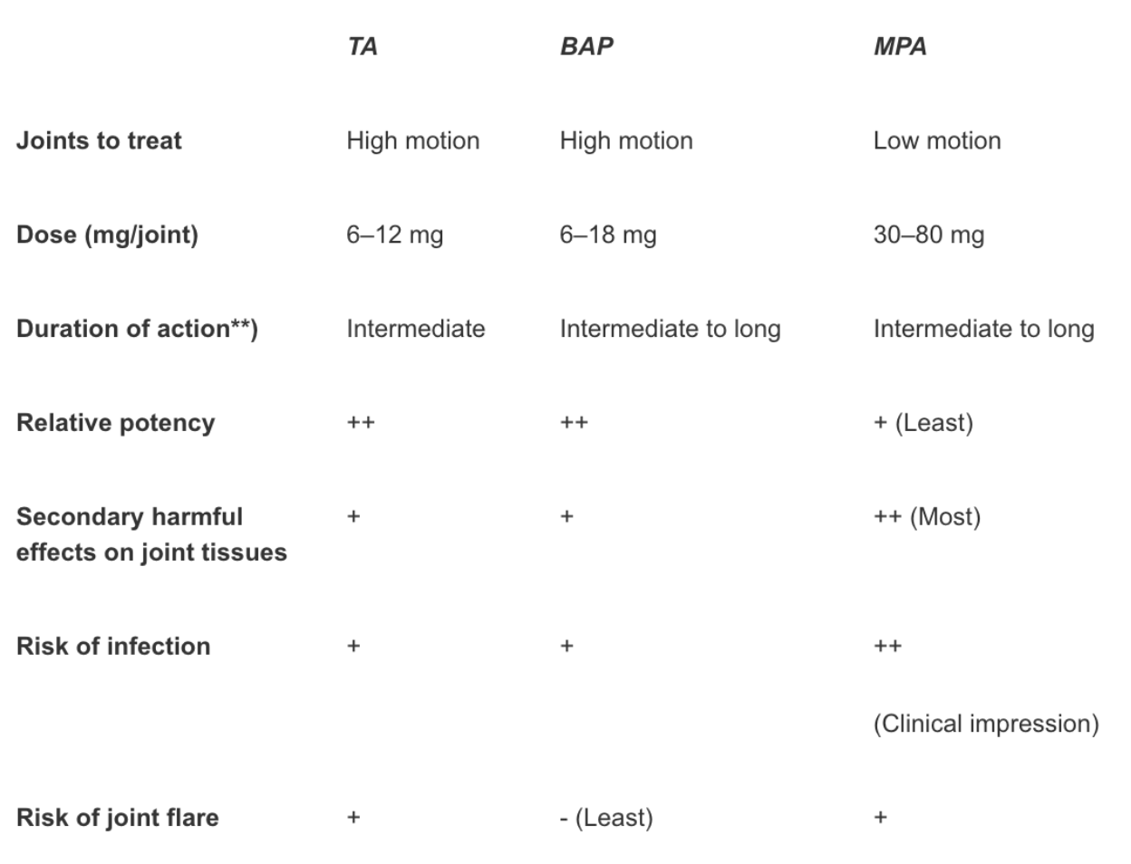
- Intra-articular (IA) corticosteroids are commonly used to treat joint problems in horses. Although a variety of corticosteroid preparations may be used intra-articularly, the 3 most commonly used drugs are methylprednisolone acetate (MPA)a, triamcinolone acetate (TA)b, and betamethasone acetate and sodium phosphate (BAP)c. The obvious clinical questions regarding IA corticosteroid use in horses are: 1) when to use which drug in what joint, 2) what is the most appropriate dose to use in different joints, 3) how can the detrimental effects of corticosteroids be minimized, and 4) how long will each drug provide clinical benefits to the horse. These and other questions regarding the clinical use of corticosteroids in horses will be addressed.
- Regarding potency, it has been estimated that BAP is more potent than TA, which is more potent than MPA. However, this is controversial and some veterinarians feel that TA is the most potent IA corticosteroid.
- A very simplistic approach in selecting an appropriate corticosteroid is to use TA or BAP in high motion joints (all joints other than the distal tarsal joints) and MPA in low motion joints such as the distal tarsal joints. However, there are exceptions to this generalization. Most joints are clipped prior to the injection, although there should be minimal problems injecting non-clipped joints if they are prepared adequately. I rarely combine antimicrobials (gentamicin or amikacin) with the corticosteroid and hyaluronan unless I feel there is an increased risk of infection associated with medicating a particular joint. A light bandage is place over the injection site, and oral phenylbutazone is given for 5 days to help prevent joint flare. Most horses are not worked for at least 1–2 weeks following injection, although an extended rest is not recommended unless the problem in the joint warrants a period of inactivity. The IA treatment may also be combined with systemic therapies such as IV hyaluronan (Legend), IM PSGAG (Adequan) or nutraceutical compounds depending on the clinical problem and finances of the client.
- The recommended dose of TA in the package insert is 6–18 mg/joint (1–3 ml). I usually do not exceed 12 mg/joint or 18–24 mg total dose.
- Negative Effects of IA Corticosteroids
- The 2 primary reasons not to utilize IA corticosteroids to treat horses with joint disease are the risk of synovial infection and the potential detrimental effects the drugs may have on articular cartilage and subchondral bone. Although infection is always possible, it is rare if good aseptic technique is used during the injection process. Combining antimicrobials with the injection should further decrease the risk of infection.
- Sheep and goats
- Dogs and cats vaccination
- https://www.evernote.com/shard/s648/sh/c892add8-43bb-49f5-a77d-910fcc131b47/aeec3acd4d55924675891a82eaf12b6b
- Clinical cases and treatments
- Foal Care
- Search any disease vet medicine
- Diseases, conditions and diagnosis of veterinary medicine diseases.
- Pharma - past Qs
- Misc topics: GP Notes
- humerus fracture in camels
- Some developed infections, stayed recumbent for more than 5 years without proper treatment and supportive care
- padded hooves, its balanced , soft and non concussionary gait?
- Clinical cases and treatments
- Bovines
- Camel Diseases
- Clinical Procedures for Reptiles read more
- Foal Care
- Interview Prep
- Parameters and interview questions
- Law No. 8 of 1995 on Practicing Veterinary Medicine source
- Article 2
- Any person who practices, either by himself or under the supervision of another, the following acts on an animal shall be deemed a practitioner of veterinary medicine
- 1 - Examining, diagnosing or assessing the progress of a disease.
- 2 - Dispensing, prescribing or giving drugs.
- 3 - Practising any medical or surgical procedure.
- 4 - Collecting a biological sample for making a medical diagnosis.
- 5 - Providing a certificate or medical report on an animal’s health.
- 6 -Conducting legitimate medical examinations or autopsies, or providing any medical reports thereon.
- Any person who practices, either by himself or under the supervision of another, the following acts on an animal shall be deemed a practitioner of veterinary medicine
- Article 3
- Veterinary medicine shall only be practiced with a licence from the Ministry. The applicant shall hold a bachelor’s degree or its equivalent in veterinary and animal surgery from a recognized college or institute.
- Article 4
- Article 14
- Veterinary doctors shall perform their work according to the highest standards of professionalism, ethics and honesty. In particular, they shall not:
- 1. Refuse to treat an animal.
- 2. Conduct tests or research on animals without the approval of the Ministry.
- 3. Combine their practice with any other activity or business.
- 4. Advertise their services in a manner inconsistent with the dignity of their profession.
- 5. Promote establishments or medical products, either directly or indirectly.
- 6. Allow the use of their name in the promotion of medicines, drugs or animal treatments, or lend their name to any commercial purpose.
- 7. Publish false information or information not documented in the doctors’ registry about their degrees, honours or type of specialization in any medical publication or anywhere on the door of their office or their home.
- Veterinary doctors shall perform their work according to the highest standards of professionalism, ethics and honesty. In particular, they shall not:
- Article 15
- Where a veterinary doctor suspects that an animal is infected with an epidemic or contagious disease, he shall immediately inform the competent veterinary medical centre and follow the decisions and instructions issued by the Ministry on epidemic or contagious diseases, whether or not such diseases are confined to animals or shared between humans and animals
- Article 2
- Cold Chain Components
- The cold chain has three main components: transport and storage of medicine, trained personnel, and efficient management procedures. All three elements must combine to ensure safe vaccine transport and storage.
- Prescription Requirement
- Controlled Drug (CD) prescription writing requirements:
- Patient name and address.
- Drug name.
- Dose (‘as directed’ on its own is not permitted)
- Formulation.
- Strength (where appropriate)
- Total quantity/dosage units of the preparation in both words and figures (for liquids, total volume in ml)
- Prescriber signature and address.
- Controlled Drug (CD) prescription writing requirements:
- Controlled drugs Classes
- The five classes of drugs are narcotics, depressants, stimulants, hallucinogens, and anabolic steroids.
- Lead Poisoning in Animals
- Lead poisoning in mammalian and avian species is characterized by neurologic disturbances, gastrointestinal upset, hematologic abnormalities, immunosuppression, infertility, and renal disease. The nature of the clinical manifestations is influenced by the dose and duration of lead exposure. Chelation therapy, cathartics, thiamine administration, and sedation are common treatment methods. In food-producing species, treatment is discouraged because of food safety concerns, the prolonged treatment period, permanent degenerative damage, and a poor prognosis. Tissue analysis for lead supported by pathologic abnormalities is essential to confirm the diagnosis.
- Acute lead poisoning is more common in young animals. The prominent clinical signs are associated with the GI and nervous systems. In cattle, clinical signs that appear within 24–48 hours of exposure include ataxia, blindness, salivation, spastic twitching of eyelids, jaw champing, bruxism, muscle tremors, and convulsions.
- Treatment and control
- Thiamine (2–4 mg/kg per day, SC) alleviates clinical manifestations and reduces tissue deposition of lead. Combined Ca-EDTA and thiamine treatment appears to produce the most beneficial response.
- Copper Poisoning in Sheep
- Acute and chronic copper poisoning (toxicosis) can affect animals in most parts of the world. Sheep are most frequently affected, although other species are also susceptible.
- Copper is used as a feed additive for pigs at 125–250 ppm; concentrations >250 ppm are potentially toxic—although as for sheep, other factors may be protective, eg, high concentrations of protein, zinc, or iron.
- Acute copper toxicosis causes severe gastroenteritis characterized by anorexia, signs of abdominal pain, diarrhea, dehydration, and shock. Hemolysis and hemoglobinuria may develop after 3 days if the animal survives the initial event. The sudden onset of clinical signs in chronic copper poisoning is associated with a hemolytic crisis.
- Infectious Bovine Rhinotracheitis (IBR)
- Infectious bovine rhinotracheitis (IBR) is a disease characterized by acute inflammation of the upper respiratory tract. BoHV-1 infection can also sporadically cause abortion in cattle.
- BoHV-1 infection in cattle is manifested as upper respiratory tract disease and disease of the reproductive tract.
- Clinical signs are influenced by the age of the animal, the dose of virus, route of infection and whether other agents are also present.
- Vaccination – The use of live vaccines is preferred above the inactivated ones because of the superior efficacy in clinical protection and more importantly in reduction of the virus circulation in newly infected animals. MSD Animal health market live (Bovilis IBR Marker Live) and inactivated (Bovilis IBR Marker Inac) vaccines.
- Malignant Catarrhal Fever
- MCF results from infection by one of several members of a group of closely related ruminant gammaherpesviruses of the Rhadinovirus genus. Although the MCF group of ruminant rhadinoviruses currently comprises approximately 10 known members, only a few are known to be pathogenic under natural conditions. The principal carriers and their viruses are sheep (ovine herpesvirus-2), wildebeest (alcelaphine herpesvirus-1), and goats (caprine herpesvirus-2). Another strain of unidentified origin has caused MCF in white-tailed deer. Virtually all clinical cases are caused by the sheep or wildebeest viruses.
- Disease course may range from peracute to chronic. Cases in deer and bison are often peracute with sudden death. Deer that survive for a few days and bison usually develop hemorrhagic diarrhea, bloody urine, and corneal opacity before expiring. High fever (106°–107°F [41°–41.5°C]) and depression are common. Other possible signs include catarrhal inflammation; erosions and mucopurulent exudation affecting the upper respiratory, ocular, and oral mucosa; swollen lymph nodes; lameness; and CNS signs (depression, trembling, hyporesponsiveness, stupor, aggressiveness, convulsions).
- Post Parturient hemoglobinuria
- FMD treatment
- Dead vaccine
- Antibiotics which do not affect the metabolism of protein
- Penicillin antibiotics
- Lumpy skin disease
- Lumpy skin disease is an infectious, eruptive, occasionally fatal disease of cattle characterized by nodules on the skin and other parts of the body. Secondary bacterial infection often aggravates the condition.
- Lumpy skin disease (LSD) is a disease of cattle and buffalo caused by a capripox virus.
- Clinical signs
- Infected cattle develop fever, lacrimation, nasal discharge, and hypersalivation, followed by the characteristic eruptions on the skin and other parts of the body in ~50% of susceptible cattle. The incubation period is 4–14 days.
- The nodules are well circumscribed, round, slightly raised, firm, and painful and involve the entire cutis and the mucosa of the GI, respiratory, and genital tracts. Nodules may develop on the muzzle and within the nasal and buccal mucous membranes. The skin nodules contain a firm, creamy-gray or yellow mass of tissue. Regional lymph nodes are swollen, and edema develops in the udder, brisket, and legs. Secondary infection sometimes occurs and causes extensive suppuration and sloughing; as a result, the animal may become extremely emaciated, and euthanasia may be warranted. In time, the nodules either regress, or necrosis of the skin results in hard, raised areas (“sit-fasts”) clearly separated from the surrounding skin. These areas slough to leave ulcers, which heal and scar.
- The disease may be confused with the less clinically important pseudo-lumpy skin disease, which is caused by a herpesvirus (bovine herpesvirus 2).
- Blood parasites pathogenesis and diagnosis
- Temprature variation > Theilleria > Babesia > Trypanosoma
- CFT + Latex Agglutination Test + CPR
- Cytology > Leukocytes + Erthrocytes
- Thrombocytopenia
- Bilirubin > 4x higher
- Globulin > acute phase proteein
- immune mediated keratitis
- Fasciola Hepatica in Ruminants
- Anthrax - Splenic fever, Siberian ulcer, Charbon, Milzbrand
- Braxy
- Photosensitisation occurs either as a primary condition following ingestion of photodynamic agents or secondary to liver damage, resulting in retention of the photosensitising agent phylloerythrin. Typical cases of photosensitization affect the muzzle and non-pigmented skin. The affected skin may ooze serum. During the later stages, the affected skin becomes dry and parchment-like and sloughs off.
- Ringworm - Dermatophytosis
- Misc Notes
- Trueperella (formerly Arcanobacterium) pyogenes is the most common isolate from lung tissue samples of adult cattle with chronic suppurative pulmonary disease at necrospy. Trueperella pyogenes is the most common isolate from lung lesions
- Misc topics: GP Notes
- Pregnancy Toxemia in Ewes and Does read
- Neonatal Intensive Care and Emergencies in Foals read - it’s important
- Diarrhea in Neonatal Ruminants read
- Diarrhea due to enterotoxigenic E coli is seen in calves <3–5 days old, rarely later. However, the age of susceptibility may be extended in the presence of other pathogens. Onset is sudden. Profuse amounts of liquid feces are passed, and the calves rapidly become depressed and recumbent. Calves may lose >12% of body weight in fluid within hours, and hypovolemic shock and death may occur in 12–24 hours. Body temperature may be increased but is commonly normal or subnormal. If fluid and electrolyte therapy is administered early, response is usually good. Disease produced by attaching and effacing E coli is seen predominantly in calves from 4 days to 2 months old and may manifest with diarrhea or primarily as dysentery with blood and mucus in the feces. The clinical course is short.
- Diarrhea due to Salmonella spp usually is not seen in calves <14 days old. It is characterized by feces that are foul smelling and contain blood, fibrin, and copious amounts of mucus.
- Diarrhea due to rotavirus, coronavirus, and other viruses usually is seen in calves 5–15 days old but can affect calves up to several months of age. Affected calves are only moderately depressed and often continue to suck or drink milk. The feces are voluminous, soft to liquid, and often contain large amounts of mucus. Diarrhea commonly persists for 3 to several days, with some cases of coronaviral diarrhea becoming chronic. Cases of viral diarrhea that are uncomplicated by other pathogens commonly respond within a few days to fluid and electrolyte therapy and adequate nutritional support.
- Cryptosporidiosis is seen in calves 5–35 days old but most commonly in the second week of life. It is characterized by persistent diarrhea that does not respond to therapy. Diarrhea due solely to Cryptosporidium spp is often mild and self-limiting, although the severity may be related to the general strength of the calf and to the intensity of challenge with the organism.
- Dietary diarrheas are seen in calves <3 weeks old and are characterized by voluminous feces of pasty to gelatinous consistency.
- Treatment and control
- Symptomatic therapy that restores hydration and acid-base- and electrolyte balance
- Calves that are recumbent, show evidence of water loss of ≥8% of their body weight, or that are unwilling to voluntarily ingest fluids orally require IV fluid therapy. These calves are usually acidotic, and the fluid and base deficits can be corrected initially by rapidly administering a hypertonic solution of sodium bicarbonate (either 500 mL of a 4.2% solution, or 250 mL of an 8.4% solution), followed by a physiologically balanced electrolyte solution administered at up to 40 mL/kg/hour until the volume deficit is corrected. Because diarrheic calves are frequently hypoglycemic, adding 25–50 g of dextrose to the IV fluids is often beneficial in the initial treatment phase. Oral electrolyte solutions should be used concurrently with and after IV fluid therapy to compensate for ongoing fluid and electrolyte losses.
- Fluid and electrolyte therapy is most important and should be started as soon as possible regardless of whether clinical evidence of dehydration has developed (clinical signs of dehydration are not apparent until the calf has lost at least 6% of its body weight in fluid). Calves still able to stand and willing and able to suck can often be treated with oral electrolyte solutions alone. Fluids for oral rehydration should promote the cotransport of sodium with glucose and amino acids and should contain sodium, glucose, glycine or alanine, potassium, and either bicarbonate or citrate or acetate as alkalinizing agents.
- Because the large majority of cases of bacteremia and septicemia in neonatal calves are associated with E coli, the chosen antibiotic should be effective against gram-negative bacteria.
- In several studies, severely affected diarrheic calves treated with NSAIDs in conjunction with fluid therapy showed fewer signs of pain, made a faster recovery, and had better weight gains in the reconvalescent period. These effects reported for several NSAIDs have been attributed to their analgesic, anti-inflammatory, antipyretic, and antisecretory properties.
- The use of drugs to reduce intestinal motility such as hyoscine-N-butylbromide or atropine is sometimes advocated, because they decrease fecal output. The literature does not provide any strong supportive evidence for or against the use of antimotility drugs.
- Intestinal gels and adsorbents, such as kaolin and pectin, are in general use, but their only established effect is to increase fecal consistency; they do not reduce the loss of water and ions.
- ostertagiosis
- Falcon diseases
- Cat and Dog toxicities and diseases
- Equine diseases
- Brucellosis is a bacterial disease caused by contact with infected animals and contaminated animal products like raw milk. In horses, brucellosis can cause lameness due to joint infection, including fistulous withers and “poll evil.” It can also cause abortions in pregnant mares, although this is less common.
- In horses, brucellosis causes fistulous withers, or “poll evil,” which is when there is inflammation and swelling in the bursa (the sac of fluid between bone and soft tissue) of ligaments. This inflammation and swelling typically affects the withers (highest part of the horse’s back) or poll (the part of the horse’s head behind the ears). The inflamed tissue might eventually burst, resulting in drainage of pus and further infection of the surrounding tissue. Brucellosis can also affect an animal’s ability to reproduce, though it rarely causes abortions in horses.
- Diseases that cause encephalitis
In the United States, mosquitoes spread several viruses that can cause encephalitis in horses, including eastern equine encephalitis (EEE), West Nile, and western equine encephalitis (WEE). Horses can be vaccinated for EEE, West Nile virus, and WEE. Venezuelan equine encephalomyelitis (VEE) can also affect horses, but it is incredibly rare in the United States. - Leptospirosis (Leptospira spp.)
Leptospirosis is a bacterial disease that can affect people and animals. Many kinds of animals can carry the bacteria in their urine, including horses.Signs in horses: In horses, leptospirosis is most often associated with eye problems, such as cloudiness or the eye or uveitis (inflammation in the eye). It can also cause other symptoms such as fever, stillbirths and abortions, and kidney failure. - Tickborne Diseases
Horses are at risk for tickborne infections, including Lyme disease, tularemia, and anaplasmosis. Horse owners should talk to a veterinarian about preventing ticks on horses. Treating horses for ticks can reduce the risk of tickborne diseases for you and your horse. For example, anaplasmosis is caused by the bacteria Anaplasma phagocytophilum. In horses, signs and symptoms can include fever; depression; lack of appetite; swelling of limbs, the underside of the chest, or abdomen; and discoloration of mucous membranes.
- Toxicology
- Common enteric diseases
- Common enteric diseases of cattle include simple indi-
gestion, rumen acidosis, parasites, coccidiosis, bovine viral diarrhea (BVD), winter
dysentery, Salmonella, paratuberculosis (Johne disease), molybdenosis (copper defi-
ciency), and malignant catarrhal fever (MCF), along with a wide range of toxicities
including a host of poisonous plants. The only disease on this list that is likely to truly
benefit from antimicrobial therapy is Salmonella enteritis; however, an argument could
be made for BVD and MCF.
- Common enteric diseases of cattle include simple indi-
Fundamental concepts
Differential diagnosis
include foot and mouth disease. Orf may cause ulcertaions on the mucous membranes but is not usually a cause of high fever or mortality. Clostridial disease may cause sudden death and fever/oedema but this is usually sporadic and in unvaccinated sheep.
- Contagious ecthyma is an infectious dermatitis of sheep and goats that affects primarily the lips of young animals.
- The primary lesion develops at the mucocutaneous junction of the lips and around erupting incisor teeth and may extend to the mucosa of the buccal cavity.
- The causal parapoxvirus is related to pseudocowpox (see Pseudocowpoxin Cattle) and bovine papular stomatitis (see Papillar Stomatitis in Large Animals). Infection occurs by contact.
- capripoxviruses cause sheep and goat pox
- There are two phases of metabolism. Phase I includes oxidation, reduction, and hydrolysis mechanisms. These reactions, catalyzed by hepatic enzymes, generally convert foreign compounds to derivatives for Phase II reactions. Products of Phase I, however, may be excreted as such, if polar solubility permits translocation. Phase II principally involves conjugation or synthesis reactions. Common conjugates include glucuronides, acetylation products, and combinations with glycine. Metabolism of xenobiotic agents seldom follows a single pathway. Usually, a fraction is excreted unchanged, and the rest is excreted or stored as metabolites.
- Bacterial Diseases
- Camel diseases
- sheep and goat diseases
- horse diseases
- General topics
Subjectwise classification
- Medicine
- Sport Medicine and Surgery
- Diagnostic Radioloy
- Clinical Pathology
- Pharmacology
- Reproduction
- General topics
- Anesthesia
- Vaccination
- Necropsy
Specie wise sub categories
- Equine
- Bovine
- Canine
- Feline
- Caprine
- Ovine
- Camelidae
- Exotics
- Exam Questions
- Difference between FMD and Bluetongue in Goats (Etiology, Epedemiology, Pathogenesis, Clinical signs, diagnosis, treatment, control)
- A lactating cow is presented with anorexia, distended juglar vein, depression, muffled heart sound. What is your diagnosis.
- Abomasal displacement is a very common condition in dairy cows. What are the types of Abomasal displacement, their prognosis and treamment.
- A sheep flock is suffering with anorexia, nasal discharge, paralysis of hind limbs, and discolouration of wool. what is your diagnosis and treatment.
- Write a note on trypanosoma in camels
- What are the types of subclinical mastitis, diagnosis and treatment?
- Write a short note on followings;
- CCPP
- Pregnancy toxemia in sheep
- Copper deficiency in sheep
- Polioecephalomalacia in camels
- ?
- Consult Page 146 includes dosages of common antibiotics in camels