For UAE Vet Exam and Qatar GP Exam - Diseases- vet medicine
Source: msdvetmanual.com
- Downer cow syndrome - Bovine Secondary Recumbency
- Hypocalcemia - Milk fever - Parturient Paresis
- Abomasal Displacement
- Hypomagnesemia - Hypomagnesemic tetany - Grass tetany - Grass staggers
- Ketosis - Acetonemia - Ketonemia
- contagious ophthalmia ASSOCIATED WITH MYCOPLASMA CONJUNCTIVAE IN SHEEP
- Pink eye - Snow blindness - Infectious Keratoconjunctivitis
- Brucellosis
- Bluetongue
- Bovine Ephemeral Fever
- Nutritional deficiencies - Nutritional Deficiency Anemia
- Copper Deficiency - Enzootic ataxia in sheep - swayback
- Salmonellosis
- Trypanosomiasis
- Cryptosporidiosis
- Fat Cow Syndrome
- Black Disease - Infectious Necrotic Hepatitis in Animals
- Rift Valley Fever in Sheep
- Pregnancy Toxemia in Sheep
- Contagious Footrot in Sheep
- Fusobacterium necrophorum, a gram-negative anaerobic bacteria, is a normal resident of manure-contaminated environments.
- Footrot is a subacute or acute necrotic infection that originates in the interdigital skin, leading to cellulitis in the digital region.
- The first sign is swelling and erythema of the soft tissues of the interdigital space and the adjacent coronary band. The inflammation extends to the pastern and fetlock.
- Benign Footrot
- The infection is confined largely to the interdigital skin, with only minimal underrunning of the adjacent horn. Clinically, benign footrot appears similar to ovine interdigital dermatitis, but D nodosus is involved—a situation that is hard to assess because culture of D nodosus is difficult and rarely done.
- Virulent Footrot
- Virulent footrot is a specific, chronic, necrotizing disease of the epidermis of the interdigital skin and hoof matrix that begins as an interdigital dermatitis and extends to involve large areas of the hoof matrix. Because the sensitive lamina and its network of capillaries are destroyed by the infection, the hoof wall (corium) loses its blood supply and anchorage to the underlying tissue and becomes detached. Footrot is extremely contagious and, under suitable conditions and susceptible genetics, morbidity may approach 100%. The infection is also rarely found in goats, deer, and cattle. The potential for genetic selection for increased resistance to footrot has been established.
- Treatment and control
- The most effective solution is 10% w/v zinc sulfate with 0.2% v/v of laundry detergent containing nonionic surfactants such as sodium lauryl sulfate. Aerosol sprays have been used in lieu of foot bathing and include zinc sulfate, tincture of iodine, tetracycline, copper sulfate, formalin, chlorine bleach, and other disinfectants. However, sprays are not as effective as foot bathing or soaking in zinc sulfate.
- The advent of long-acting antibiotics used in combination with topical foot treatments has improved recovery and reduced carrier animals. Parenteral treatment using a long-acting oxytetracycline at 13.6 mg/lb gives a duration of effect in cattle of 7–8 days and probably a similar duration of effect in sheep.
- Camel Pox Virus Camelpox (CMLV)
- CCPP
- Mycoplasma capricolum capripneumoniae (Mycoplasma biotype F38) is the causative agent. It appears to be transmitted by infective aerosol.
- Contagious Caprine Pleuropneumonia (CCPP) is a highly contagious infectious disease of goats caused by the Mycoplasma mycoides capri and Mycoplasma F38 bacteria. CCPP causes inflammation of the lungs and accumulation of fluid in the chest cavity. Damaged lung tissue can harden and adhere to the chest wall, which interferes with effective respiration and causes the goat to die from lack of oxygen. Mortality rates can reach 100 percent.
- Pneumonia and pleuropneumonia can be caused by other mycoplasmas, including M mycoides capri. Taxonomic change means this subspecies also includes M mycoides mycoides large colony type. Morbidity and mortality rates are generally lower with M mycoides capri, and joint and udder infections may also be seen.
- Contagious Bovine Pleuropneumonia
- Contagious bovine plueuropneumonia is highly contagious and generally accompanied by pleurisy. The causal organism is Mycoplasma mycoides mycoides small colony type.
- In acute cases, signs include fever up to 107°F (41.5°C); anorexia; and painful, difficult breathing. In hot climates, the animal often stands by itself in the shade, its head lowered and extended, its back slightly arched, and its elbows turned out. Percussion of the chest is painful; respiration is rapid, shallow, and abdominal.
- Nairobi sheep disease (NSD)
- Nairobi sheep disease (NSD) is a tickborne viral disease of sheep and goats characterized by fever and hemorrhagic gastroenteritis, abortion, and high mortality
- Foot and Mouth Disease
- Foot-and-mouth disease (FMD) is a highly transmissible disease caused by infection with an Aphthovirus, a member of the family Picornaviridae. There are 7 serotypes of the virus, termed: A, O, C, Asia 1, and SAT (Southern African Territories) 1, 2, and 3. Further diversity is found between strains within each serotype. The virus primarily affects cloven-hoofed animals of the order Artiodactyla. Livestock hosts include cattle, pigs, sheep, and goats. FMD virus has also been reported to affect >70 species of wild artiodactyls, including African buffalo, bison, giraffes, camels, and several species of deer and antelope.
- FMD is characterized by fever and vesicles in the mouth and on the muzzle, teats, and feet of animals and is spread through contact with infected animals or their excretions.
- The primary site of infection and replication of FMD virus is in the mucosa of the pharynx. The virus may also enter through skin lesions or the GI tract. Once distributed throughout the lymphatic system, the virus replicates in the epithelium of the mouth, muzzle, teats, feet, and areas of damaged skin (eg, knees and hocks of pigs). Vesicles then develop and rupture within 48 hours.
- Clinical signs in cattle include fever of ~40°C, followed by vesicular lesion development on the tongue, hard palate, dental pad, lips, gums, muzzle, coronary band, interdigital cleft, and teats in lactating cows.
- Vesicles (fluid filled blisters) on the tongue, dental pad and hard palate quickly rupture leaving shallow ulceration with shreds of mucosa at the periphery. The underlying tissues are reddened and painful.
- Ruptured vesicles (fluid-filled blisters) on the tongue revealing reddened and painful ulceration with shreds of mucosa at the periphery.
- Vesicles may be present on the teats and at the coronary band (top of the hoof). The latter can become secondarily infected, causing lameness.
- There are no ocular or nasal discharges. During the acute phase of disease, there is marked weight loss and milk yield reduction. Foot lesions often become secondarily infected and animals are very lame and often may be reluctant to rise.
- Rabies
- Rabies is caused by lyssaviruses in the Rhabdovirus family, Lyssavirus genus.
- Rabies is an acute, progressive encephalomyelitis caused by lyssaviruses. It occurs worldwide in mammals, with dogs, bats, and wild carnivores the principle reservoirs. Typical signs include acute behavioral change and progressive paralysis. The disease is fatal once clinical signs appear, but treatment with local wound care, immune globulin, and vaccination can prevent disease in humans following exposure. Vaccines are available for domestic animals, wildlife, and people to prevent rabies and help control spread in reservoir populations.
- Three forms are described and include 1) cerebral or furious: aggressive behavior, photophobia, hyperesthesia, straining, and convulsions), 2) brainstem or dumb form: depression and dementia with ataxia, excessive drooling and pharyngeal paralysis and 3) spinal cord or paralytic form: progressive ascending paralysis. Common cattle signs in order of most common are salivation, bellowing, aggressiveness, paresis or paralysis and straining.
- Clinical signs
- Clinical signs of rabies are rarely definitive. Rabid animals of all species usually exhibit typical signs of CNS disturbance, with minor variations among species. **The most reliable signs, regardless of species, are acute behavioral changes and unexplained progressive paralysis. **Behavioral changes may include sudden anorexia, signs of apprehension or nervousness, irritability, and hyperexcitability (including priapism). The animal may seek solitude. Ataxia, altered phonation, and changes in temperament are apparent. Uncharacteristic aggressiveness may develop—a normally docile animal may suddenly become vicious. Commonly, rabid wild animals may lose their fear of people, and normally nocturnal species may be seen wandering about during the daytime.
- The clinical course may be divided into three general phases—prodromal, acute excitative, and paralytic/endstage. However, this division is of limited practical value because of the variability of signs and the irregular lengths of the phases. During the prodromal period, which lasts ~1–3 days, animals show only vague nonspecific signs, which intensify rapidly. The disease progresses rapidly after the onset of paralysis, and death is virtually certain a few days thereafter. Some animals die rapidly without marked clinical signs.
- Hardware disease/Traumatic reticuloperitonitis
- Anaplasmosis / Yellow fever/ yellow bag
- Babesiosis
- Theileriosis
- Parrasitic diseases in sheep
- Clostridial Diseases
- Dummy Syndrome - Dummy Foal
- Progressive ataxia (PA) of Charolais cattle
- Progressive ataxia (PA) of Charolais cattle is an inherited neurodegenerative disease affecting the hind limbs that can gradually progress until the affected animal is unable to stand.
- Progressive ataxia is a fatal hereditary defect (lethal mutant) resulting from irreversible changes in the brain and spinal cord. The peripheral nerves do not show any changes. This hereditary defect is insidious because no signs of this disease are seen in calves. The disease usually begins insidiously at 18 to 24 months of age with weakness in the hind legs and crossing of the legs
- Progressive ataxia (PA) of Charolais cattle is characterized by onset of unsteady gait and stiff hind limbs with gradual worsening of the condition that results in an inability to stand and permanent recumbency (lying down). Other signs of the disease include head bobbing when excited and, in females, irregular pulsatile urination.
- SHEEP MITES: biology, prevention and control. Sheep scab, sheep mange. Psoroptes, Sarcoptes, Chorioptes, Psorergates, Demodex
- Mites infest sheep worldwide. The most important parasitic mite species of sheep are:
- Psoroptes ovis that causes psoroptic mange, also called sheep scab: worldwide
- Sarcoptes scabiei var.*** ovis*** that causes sarcoptic mange, also called scabies: worldwide
- Chorioptes ovis that causes chorioptic mange, also called leg mite, foot scab: worldwide
- *Psorergates ovis____,* responsible for psorergatic mange, also called itch mite. Especially in Australia, New Zealand, South Africa, North and South America.
- Demodex ovis, responsible for sheep demodectic mange can have local importance. In most cases it causes no clinical symptoms and has little or no economic impact on sheep flocks.
- Mites infest sheep worldwide. The most important parasitic mite species of sheep are:
- Sarcoptic Mange
- In sheep, Sarcoptes spp. is an important cause of mange that leads to itch, dermatitis and intense pruritis due to which animals loose much of the grazing time and hence loose general body condition.
- Sarcoptes scabiei var ovis infests sheep, and S scabiei var caprae infests goats, throughout the world. However, S scabiei var ovis is rare in the USA. This mite infests nonwooly skin, usually on the head and face. Typical of scabies, lesions manifest with formation of crusts and intense pruritus. Affected animals have decreased reproduction, meat gain, and milk yield. In goats, S scabiei var caprae is responsible for a generalized skin condition characterized by marked hyperkeratosis. Lesions start usually on the head and neck and can extend to the inner thighs, hocks, brisket, ventral abdomen, and axillary region. Both S scabiei var ovis and S scabiei var caprae are zoonotic. Consistent with other animal variants of Sarcoptes, zoonoses are initiated from direct contact with infested animals but are self-limiting infestations.
- Microscopic examination of deep skin scrapping under 10x and 40x showing presence of
Sarcoptes spp. mange mite - Treatment and control
- Hot lime sulfur spray or dip is labeled for use against sarcoptic, psoroptic, and chorioptic mites in sheep. Treatment should be repeated every 12 days if needed. Certain formulations of permethrin sprays are labeled for mange in sheep and goats.
- Ivermectin @ 200 mcg/kg body weight
sub cutaneous (S/C) at weekly interval along with topical application and supportive
therapy. Improvement in animal condition was observed from 10 days post treatment with
full recovery within 20-25 days. Supportive therapy along with topical application of
ointment was helpful in fast healing of mange lesions.
- Chorioptic Mange
- Chorioptes bovis infests sheep and goats worldwide. Prevalence of C bovis is more common in rams than ewes or lambs. Infestation of C bovis on goats is fairly common, with most of a herd infested. Distribution of lesions is the same as that in cattle, with papules and crusts seen on the feet and legs. Most sheep are subclinically infested with C bovis. However, C bovis can cause exudative dermatitis on the lower legs and scrota of rams (scrotal mange). Semen quality may be affected, presumably due to increased temperature of infested scrota.
- Demodectic Mange (Ovine Demodicosis, Caprine Demodicosis)
- Demodex ovis infests sheep, and D caprae infests goats. Demodectic mange in sheep is not common, whereas D caprae are relatively common in goats. Lesions are similar to those in cattle. In goats, nonpruritic papules and nodules develop, especially over the face, neck, shoulders, and sides or udder. Demodectic mange in goats occurs most commonly in kids, pregnant does, and dairy goats. The nodules contain a thick, waxy, grayish material that can be easily expressed; mites can be found in this exudate. The disease can become chronic. Historically, in some cases, localized lesions in goats have been managed by incision, expression, and infusion with Lugol’s iodine or rotenone in alcohol (1:3). This practice should not be continued or condoned. Rotenone, a plant-derived ketone once a popular pesticide and ectoparasiticide approved for use in organic farming, is now available only as a piscicide in the USA and Canada.
- Psoroptic Mange (Sheep Scab, Ear Mange):
- Psoroptes ovis is a highly contagious and severe infestation of sheep. This mite has been eradicated from sheep in Canada, New Zealand, and the USA. However, sheep scab persists in many countries, including some in Europe. Intense pruritus leads to large, scaly, crusted lesions that develop in more densely haired or woolly parts of the body. Lesions begin on the back and side but may become generalized and cover a large portion of the body. Animals bite, lick, and scratch in response to the pruritus, which results in wool loss and secondary bacterial infection. If affected sheep are not treated, infested animals may become emaciated and anemic and possibly die.
- Psoroptic mange (ear mange) in goats and sheep is caused by P cuniculi, which is likely a variant of P ovis. P cuniculi typically infests the ears of goats but can spread to the head, neck, and body. Infestation of P cuniculi in goats can be common, with 80%–90% of a herd infested. Disease can range from subclinical to scaling, crusting, inflammation, alopecia, ear scratching, head shaking, and rubbing of ears and head to alleviate irritation. Although the course is chronic, the prognosis is good with appropriate treatment.
- Polioencephalomalacia - or Cerebrocortical necrosis (PEM or CCN)
- Gid - Coenurosis - __ Staggers - Sturdy
- Mycotic Abortion in Cattle
- Fungal placentitis due to Aspergillus sp (septated fungi, 60%–80% of cases), or to Mucor sp, Absidia, Rhizopus sp, and a few other nonseptated fungi, is an important cause of sporadic bovine abortion. Abortions occur from 4 months to term and are most common in winter. It is believed the fungi gain entry through the oral or respiratory tracts and travel hematogenously to the placenta. Placentitis is severe and necrotizing. Cotyledons are enlarged and necrotic with turned-in margins. The intercotyledonary area is thickened and leathery. Adventitious placentation is common. The fetus seldom is autolyzed, although it may be dehydrated; ~30% have gray ringworm-like skin lesions principally involving the head and shoulders. The diagnosis is based on the presence of fungal hyphae associated with necrotizing placentitis, dermatitis, or pneumonia. Fungi can also be isolated from the stomach contents, placenta, and skin lesions. Isolation must be correlated with microscopic and gross lesions to exclude contamination after abortion.
- Others; Vaccination protocols, common drugs mechanisms,
- Deficiencies -B1, Selenium
- Copper Deficiency
- Selenium and Vitamin E deficiency - White Muscle Disease
- Cobalt deficiency
- anemia, poor coat,
- Improved growth following vitamin B12 injections in a controlled study is the best means of establishing the diagnosis.
- Principles of Biosecurity of Animals, source
- all procedures implemented to reduce the risk and consequence of infection with a disease-causing agent. This broad definition recognizes that disease is a complex interaction between the host, the disease-causing agent, and the environment. Biosecurity can be considered in terms of individual animals or populations of animals (flocks or herds), economic entities (production facilities or companies), or geographic regions (counties, states, countries, or continents), thus facilitating compartmentalization for trade purposes. Importantly, it addresses strategies for both disease prevention (eradication) and control (limiting the consequence of infection).
- Biosecurity is the implementation of measures that reduce the risk of the introduction and spread of disease agents; it requires the adoption of a set of attitudes and behaviors by people to reduce risk in all activities involving domestic, captive/exotic, and wild animals and their products (FAO/OIE/World Bank, 2008).
- Bioexclusion centers on the prevention of disease introduction and relies on external biosecurity practices. In contrast, biocontainment centers on preventing the spread of disease within a farm or group of animals, or to other farms or groups of animals, and relies on implementation of internal biosecurity practices.
- Thus, disease control and prevention relies on the interrelated processes of bioexclusion, surveillance, and biocontainment.
- Disease Transmission in Biosecurity
- Understanding disease transmission is central to designing proper biosecurity protocols. Diseases can be transmitted in many ways, and direct animal-to-animal contact as well as contact with contaminated fomites are some of the most common transmission routes. Contaminated semen and natural mating can be sources of sexually transmitted diseases.
- Many fomites (inanimate objects) act as carriers of disease-causing agents. Survival of agents on fomites may depend on the composition of the particular fomite and how easily it can be disinfected. Examples of fomites considered high risk include trailers, vehicles, maintenance and repair tools, boxes, materials used to remove dead animals, loading chutes, etc.
- Vectors are also capable of transmitting diseases; among the most significant vectors are birds, bats, rodents, feral and wild animals, stray and domestic animals, and insects.
- Air can be a source of diseases, particularly in areas of high animal density. Contaminated water and feed and consumption of contaminated, raw, untreated animal products have also been implicated in disease transmission. Manure, bedding, and carcasses can also be a source of agents unless disposed of properly. Lastly, people can act as both mechanical and biologic vectors, and training and awareness of personnel working with animals is necessary for proper implementation of biosecurity programs.
- Disease Prevention in Biosecurity of Animals
- Disease prevention depends on 1) stringent bioexclusion to avoid contact between the disease-causing agent and the host, 2) early detection of a breach in biosecurity through vigilant surveillance, and 3) rapid implementation of a ruthless biocontainment policy. This is feasible only if there is an effective way to detect infection; containing the infection through slaughter or other means, clean-out, and disinfection; and preventing dissemination of the disease-causing agent. Eradication is reserved for those diseases that pose a dire public health threat, that have a devastating effect on animal performance, or that severely compromise end-product quality. Elimination of diseases without a regulatory framework is common in food animals if these diseases are economically significant and their elimination is advantageous for the producers.
- Disease Control in Biosecurity of Animals
- In disease control strategies, the emphasis shifts from preventing disease to reducing its consequence or economic impact. Prevalence data are now used primarily to assess the level of protection and challenge, not merely the presence or absence of disease. Although biosecurity still relies on principles of prevention, disease-control programs focus more on limiting the extent and consequence of exposure. Many biosecurity measures aimed at preventing or eradicating epidemic disease also produce beneficial by-products, such as establishment of a firm foundation for control of erosive/endemic diseases and enhancement of host resistance through immunization.
- The Three Levels of Biosecurity of Animals source
- Conceptual Biosecurity of Animals
- primary level, location of the facility
- Structural Biosecurity of Animals
- secondary level, physical barriers, showers, construction
- Procedural Biosecurity of Animals
- Tertiary level , procedures according to the situation and diseases
- Conceptual Biosecurity of Animals
- Management - Breeding, nutrition, /Repeat breeding
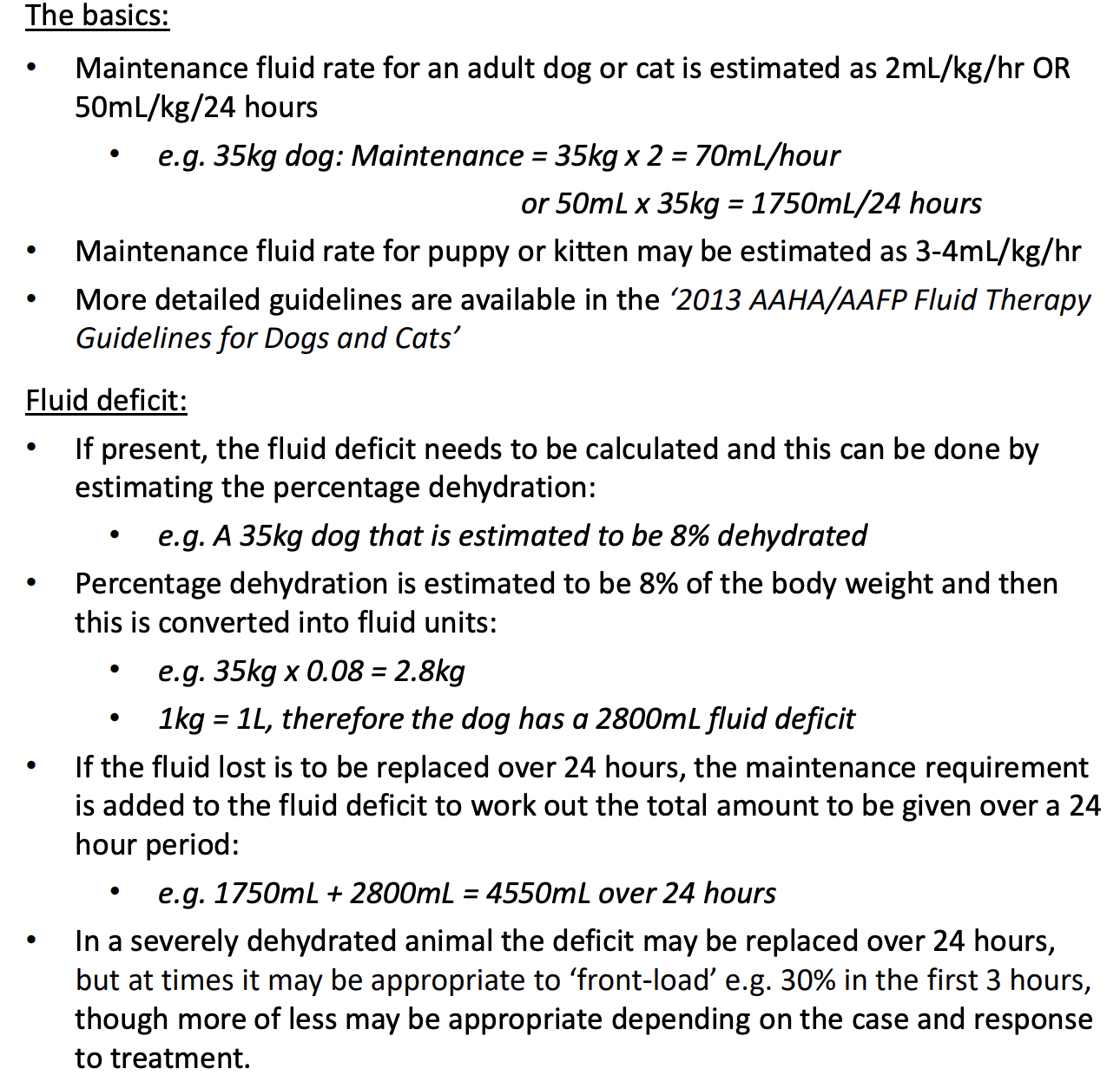
- Fluid Therapy
- Calculation
- Calculation
Overview
- downer cow syndrome is a complication of periparturient hypocalcemia. Recumbency in cattle is caused by numerous metabolic, traumatic, infectious, degenerative, and toxic disorders. If treatment of the underlying cause of recumbency is not successful and cattle are unable to rise for >24 hr after initial recumbency, they may develop a secondary recumbency from pressure damage to muscles and nerves, often termed “downer cow syndrome. Calving paralysis after dystocia may also result in recumbency due to traumatic injury to tissues and nerves inside the pelvic cavity. Regardless of the initial cause of recumbency, all cattle develop pressure-induced damage to muscles and nerves of the pelvic limbs, especially when lying on a hard surface. The hindlimb muscles of the leg the animal is lying on are compressed between the bones and the skin by the physical pressure from the weight of the recumbent cow. The positioning of the hindlimbs may indicate the cause of the recumbency. Limbs splayed out behind the animal may indicate obturator nerve paresis or paralysis, hip dislocation, or fracture of the femur or tibia. Fracture should be suspected whenever the upper limb is extended sideways in such a manner that a crease is formed in the skin.
Diagnosis and differentials
- Blood samples are not usually taken when treating routine cases of hypocalcemia. However, hypocalcemia, hypophosphatemia, and hypokalemia should be assumed to be present in all recumbent cattle, and determination of the biochemical status of cattle unresponsive to calcium therapy frequently helps guide treatment and prognosis. Hypokalemia and hypophosphatemia are commonly quoted causes of creeper cows (cows able to crawl but unable to stand). Alert downer cows may have normal serum concentrations of calcium, potassium, magnesium, and phosphorus. Downer cows have increased serum CK, AST, and LDH; cows that do not recover have higher serum AST and CK activities than cows that do recover. Increased serum CK activity is a specific indicator of muscle damage; however, CK activity peaks shortly after the start of muscle damage and declines noticeably within 4 hr. For this reason, increased serum AST activity is the best prognostic indicator in recumbent cattle, with higher AST activities indicating a poorer prognosis. In cattle with severe muscle damage, the urine may contain myoglobin as well as higher than normal concentrations of protein.
Treatment and control
- Downer cows are often hypocalcemic. If an apparently hypocalcemic cow does not respond to calcium therapy, potassium, phosphorus, and magnesium should be given as additional treatments pending results of laboratory tests. Monitoring blood mineral status is an important part of downer cow management.